Antenatal Discovery of Bilobed Placenta Helped Manage Third Stage of Labor: A Case Report
Bilobed placenta, also known as the bipartite or bilobate or duplex placenta is the placental morphological variation in which the placenta is separated into nearly equal size discs. The incidence of occurrence is less. Women age more than 35 years, multigravidas, history of infertility treated are more prone to have a bilobed placenta. The Bilobed placenta is associated with an increased incidence of first-trimester bleeding, placenta previa, adherent placenta, vasa previa and postpartum hemorrhage secondary to retained placenta. Here we present a case report of bilobed placenta diagnosed at 33 weeks scan. Prenatal diagnosis of bilobed placenta alerts the obstetrician for appropriate planning, prompt recognition, and treatment of complications.
Keywords:Bilobed Placenta; Bipartite Placenta; Bilobate Placenta; Duplex Placenta; Retained Placenta
The placenta is a fetal organ consisting of an umbilical cord, membranes (chorion and amnion), and parenchyma. The normal placenta is round to oval in shape. In contrast to the normal architecture described, placentas may infrequently form as separate, nearly equally sized discs. This is a bilobed placenta, which is also known as bipartite placenta or placenta duplex or bilobate placenta [1]. In these, the cord inserts between the two placental lobes-either into a connecting chorionic bridge or into intervening membranes. If more than two lobes are present, it is termed a trilobed, four-lobed and so on. The Bilobed placenta can be identified in the ultrasound with its characteristic sonographic appearance. But it is rare [2]. To my knowledge, there are only a few case reports that have been published.
A 36- year-old multigravida, gravida 4, parity 2, living 2, abortion 1, a booked patient in our regional hospital with regular antenatal visits. Her last pregnancy had to be terminated at 21 weeks due to the congenital anomaly (gastroschisis). Due to this reason, her antenatal care in this pregnancy was extra cautious. Multiple ultrasound scans were done from various centers. Her 20 weeks anomaly scan was normal with the placental location being on the posterior site and the lower margin being 4.4cm away from the internal Os. In 33 weeks scan, her placental impression was the bilobed placenta, mentioning two separate placental lobes on the anterior and posterior fundal wall of the uterus. Cord insertion was noted in between placentas, eccentrically in the posterior lobe. Thereafter in subsequent scans; the fetal growth was adequate, same placental findings, normal amniotic fluid index and normal umbilical artery doppler.
She was then well counseled regarding her placental features and the risk of having intrauterine growth restriction fetus. Increase incidence of postpartum hemorrhage due to retained placental tissue. She was advised to keep donors ready all the time. Mode of delivery was planned vaginally and if she has retained placenta may need exploration in the operation theatre.
She came at 39 weeks with the complaint of the ruptured membrane just prior to hospital arrival. She had a moderate meconium stained liquor. Her per vaginal examination BISHOP score was 6. Syntocin induction was started. She delivered vaginally with intact perineum, healthy female baby, weighing 2950gm. Her 3rd stage of the labor was managed carefully. Injection syntocin 10 unit intramuscular was given. The Placenta was delivered by controlled cord traction under the guidance of the ultrasound. Traction force was minimal. We could see the placental separation both anterior and posterior slowly and completely. Estimated Total blood loss was less than 150mL.
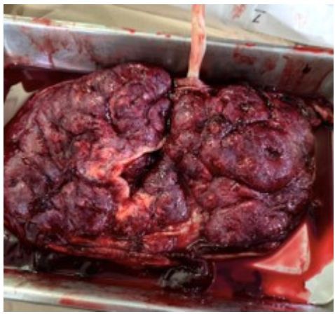
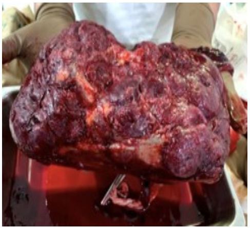
Placental morphology was explored. There were two separate almost equal lobes connected by the fetal membrane in the central. The Parenchymal separation was noted at the cord insertion site. The cord was inserted into the chorionic bridge between lobes. The placenta weighted 800gm (Figure 1a,1b and 2).
She was discharged 24hr post-delivery. Her hospital stay and postnatal period were uneventful.
Several theories have been advanced to explain the formation of the bilobate placenta. Benirschke and Driscoll favour trophotropism, the abnormal lateral growth of the placenta [3]. Torpin and his colleagues presented convincing evidence that bilobed placenta results from unusually superficial implantation of the fertilized ovum, thus enabling it to adhere and develop on both walls [4,5]. The bilobed placenta is also thought to result from localized atrophy as a result of poor decidualization and vascularisation in a part of the uterus [6].
In Torpin’s series of 4098 placentas, 8.6 percent were termed bilobate, ‘Which included varying degrees of separation of the two lobes of the placenta’ [7]. Among them, 21.5 percent showed division into two almost equal lobes.
In a study done by Fujikura et al. among white women at the Boston Hospital, the incidence of bilobed placenta reported was 4.2 percent (366 of 8,505) of placentas [1]. Women age more than 35 years, multigravidas, history of infertility was more numerous in the bilobed group. There were also increased incidence of first-trimester bleeding, placenta previa, and adherent placenta in the bilobed group. Despite this bilobed placenta was not found associated with low birth weight and neonatal death.
Other causes of the bilobed placenta may be implantation in areas of decreased blood supply, over uterine leiomyoma, previous uterine scars and implantation over the cervical Os [2]. When the implantation of the bilobed placenta is over the cervical Os it is linked with placenta previa and vasa previa [8]. There are few case reports of the bilobed placenta with vasa previa [9]. This is important because vasa previa is linked with severe neonatal morbidity and mortality.
The case presented here didn’t have any of the above mentioned complications. The antepartum identification of the placental morphology alerted the obstetrician. The case was also notified to the midwives in the labor room. An Anticipated complication of delivery was discussed. Her delivery was conducted by the obstetrician.
In a gross morphologic examination in ultrasound, it is possible to see two separate portions of the placenta: roughly equal in size. It is important not to confuse this with the placenta that covers two major aspects of the uterine cavity. In the latter case, there is a fold of connecting placenta tissue. Angtuaco described their ultrasound finding of the bilobed placenta as: A placenta covering both anterior and posterior uterine wall with a distinct point of separation dividing it into roughly equal two halves. At the point of apparent separation, the placental substance tapered to a membranous attachment [10].
In a case report by Ulrike, the bilobed placenta was identified as early as 18 weeks [11]. Others have demonstrated that it can be diagnosed as early as 26 weeks gestation [2,12,13]. In our case, we missed to identify at 20 weeks and was only identified at 33 weeks. There were no scans done in between to rule out if it could have been identified earlier in that period of time, as the patient had normal monthly antenatal checkups which were not suggestive of any placental anomaly. In our hospital after 20 weeks anomaly scan, we routinely do scans on 33 weeks for fetal well being, placental location, amniotic fluid index.
Ultrasonography has become the primary imaging method in the placental assessment. Evaluation of the placental location, maturity, a cord is must in most obstetrics scans. Early detection of placental anomaly underlies the importance of ultrasound in obstetrics. Prenatal diagnosis of the bilobed placenta in scans alerts the obstetrician and helps them appropriate planning, prompt recognition, and treatment of complications associated with it. However, the literature on this topic is scanty. More studies are require to see how early it can be detected, with its roles in fetal growth, antepartum and postpartum complication.


