“Infection of Piglets with the Porcine Respiratory and Reproductive Syndrome Virus (PRRSV): A Morphological Study
Together 28 piglets (aged over 2 months) were infected with 105 TCID50 of porcine respiratory and reproductive syndrome virus (PRRSV) into both nasal nostrils using a volume of 2x150 (i.e. 300) microliter inoculum. In addition, 9 piglets served as uninfected controls. On day 11 post-infection, tissue samples from tonsilar area, each lung lobe, spleen and liver were taken from 12 sacrificed animals. By day 18, another 16 piglets have been authopsied for tissue sampling. At both intervals, also blood samples were taken for serological examination. For histological examination, the organ samples were fixed in neutral formalin, processed and embedded into paraffin. Sections were stained either for standard histology, or treated with immunohistochemical staining reagents. A commercial monospecific antiserum against the PRRSV N-protein was applied in the first layer in combination with an alkaline phosphatase labelled second antibody applied in second layer. An additional slide was treated with the second antibody only as staining control. In 4 out of 9 uninfected (negative control) piglets a slight focal thickening of the peri-bronchial connective tissue and/or of the inter-alveolar septa was noted (referred to as mild non-specific interstitial infiltrate, MNSII). The MNSII could be clearly distinguished from usual interstitial pneumonia (UIP) as detected in the lungs of 23 out of 28 infected animals (82 %). In the latter, the inter-alveolar septa revealed more widespread mononuclear cell (mainly lymphocyte) infiltration which occasionally reached an extensive intensity. As a rule, the N-protein was found in the bronchial ciliary epithelium cells of nearly all the piglets who developed UIP (21 out of 28, 75 %), along with less frequently positive squamous epithelium at pharyngeal and/or tonsilar areas (13/28, 46 %).
Keywords:Porcine Respiratory and Reproductive System Virus (PRRSV); Usual Interstitial Pneumonia (UIP); N-antigen Detection
The porcine respiratory and reproduction syndrome virus (PRRSV) forms small, enveloped particles (50-65 nm in diameter) harboring a relatively long (approximately 15 kb in size) single strand RNA genome [2]. The viral RNA (vRNA) is a positive-sense molecule with a terminal cap at 5´-end and a poly-A repeat at 3´-end [3]. In the course of virus replication, the vRNA is copied as whole, via a full length negative-strand RNA intermediate. The vRNA sequence has 2 (two) long open reading frames (called OR- F1a and ORF1b), which together comprise about 75% of the total sequence [4]. This portion of the genome specifies 14 non-struc- tural proteins (nsps) which are formed by cleavage of the both translated polyproteins. Of special importance are, for example, two non-structural proteins (nsp9 and nsp12), which function as vRNA replicase, also termed RNA-dependent RNA polymerase (RdRp) [5]. The rest of the genome encodes 7 structural proteins, out of which 5 are glycoproteins (designated GP2a/Gp2, GP2b/E, GP3, GP4 and GP5) along with the M (membrane) protein and the N nucleoprotein [6].
Regarding to the structure of vRNA, the PRRSV has been classified as a member of family Arteriviridae (order Nidovirales), along with the equine arteriitis virus and the lactate dehydrogenase elevating virus of mice [7]. In the course of vRNA replication, a total length (genomic) minus strand is generated, which serves as template for the synthesis of new vRNA molecules. During viral mRNA synthesis, the negative sense RNA sequence is being formed first; then a set of positive sense nested subgenomic (sg) RNA molecules is transcribed. Finally, a full set of minus sense subgenomic (sg) RNAs is formed, which becomes a template for the synthesis of functional positive sense sg mRNAs [7]. Both strands are complementary to each other; the coterminal 3´-ends are equipped with a common leader sequence at their 5´-ends [8, 9]. The viral genome reveals several (but at least two) conserved transcription regulatory sequences (TRS), which are located either in the front of ORF1a (encoding the structural protein GP2a) or ORF2a (encoding the envelope glycoprotein Gp2b/E).
The classical PRRSV strains which were isolated in the US (VR2332) and/or in Europe (Lelystadt) differ at both, by serological as well as genome examinations [10]. Experimental infection with the PRRSV isolates can be lethal in newborn and/or 3-week-old piglets. A key event of infection process is the involvement of porcine alveolar macrophages, which are the most important virus target mediating virus spread [11]. To date, at least two macrophage surface molecules are known as entry mediators: the siglec sialoadhesin and a scavenger receptor CD163 [12]. The PRRSV induced pneumonia is characterized by thickening of inter-alve- olar septa due to infiltration with macrophages and by the presence of occasional inflammation and cell debris within the alveoli itself [13]. Also alveolar pneumocytes of type II may be found PRRSV antigen positive along with the hyperplasia of peribronchial lymphatic tissue [14]. The severity of lung lesions may vary from relatively mild to quite extensive. The viral genotypes can differ in their pathogenicity, namely the Type 2 North American PRRSV induces more severe respiratory disease than type 1 European virus. Nevertheless, mild thickening of interalveolar septi with focal thickening of inter-alveolar septa in combination with slight infiltration of peri-bronchial connective tissue (referred to as mild non-specific interstitial infiltrate, MNSII), was occasionally seen in a proportion of non-infected control piglets can be mistaken and interpreted as unrelated to PRRSV infection [15]. In this paper we describe the correlation of the lung lesions as seen at histological examination in comparision to the immunohistochemical de- tection of viral nucleocapsid (N)-protein along with the results of serological tests for given N-protein antibodies. Detection of the N-protein has been chosen, since it is the most abundant structural protein of the virion [16]. Though the corresponding antibody does not neutralise the virus, it is highly immunogenic. Therefore, the detection of N-antibody is a regular part of routine diag- nostic assays. We have decided to elaborate a simple N-antibody detection technic in a piglet model. To be sure that the antibodies tested have been produced in correctly infected animals, we decided analyse the virus distribution in them.
Virus. A North-American strain was cultured on the MARC-145 cell line; its titer end point (TCID50) was evaluated using a 96-well plate as described by Zhao et al. (2014)[17] and/or Ramakrishnan (2016) [18].
Animals. The piglets used were coming from a regular Hungarian breed; they were removed from their sows aa soon as manual feed-d ing has become possible. Out of a total of 37 pigs, 28 animals were inoculated into both nostrils with 105 TCID50 of above mentioned US (VR2332) strain administered in a volume of 300µl culture supernatant. The nine (9) control animals were inoculated with a virus free culture medium and then kept under careful isolation conditions to avoid any contact with the infected ones.
Specimen sampling and histological examination. At given intervals post-infection, the animals were succumbed. Blood was drawn to obtain serum, whole the tissue samples (coming from each lung lobe, from both tonsils including adjacent paryngeal area, from spleen and liver) were removed and immediately immersed into 10% neutral formalin for 24hr. Fixed tissue samples were rinsed in phosphate buffer, dehydrated in a series of corresponding reagents and embeded into paraffin as described Szeredi et al. (2006)[19] and/or Stipkovits et al. (2012) [20]. Next, the sections were stained either by classical hematoxylin and eosin (HE) and/or treated by immunohistochemical reagents, namely a commercial anti-PRRSV antibody to N protein (a mixture. of. SDOW-17 and SR-30) in the first layer. It has been purchased from 4rtilab and mixed for use in an equal 1:1 ratio.
ELISA titer meassurements. The specific antibody levels (serum class IgG) against the N-antigen of PRRSV were determined using the INgezim PRRS 2.0 ELISA kit (purchased from Eurofins) strictly following the procedure recommended by the manufacturer.
Saliva collection. The pooled oral fluid was collected from each animal separately using the Civtest suis oral fluid rope IDEXX. Ob- tained saliva samples we examined for the presence of class IgA specific antibody to the N-antigen of PRRSV. The antibody test was performed with the Oral Fluids (IDEXX PRRS OF) kit as recommended in the manufacturer’ s manual.
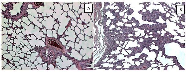
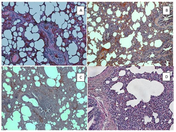
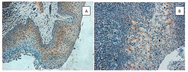
As documented in Figure 1A the interalveolar septi in normal lung tissue are very thin in order to ensure the diffusion of oxygen into blood capillaries, where the erythrocytes circulate. Occasionally, a few mononuclear cells (mainly lymphocytes) might be seen in the peribronchial connective tissue. Surprisingly, in 4 out of 9 uninfected (control) piglets, a slight focal thickening of interalveolar septi was noted along with the accumulation of relatively few interstitial infiltrate consisting of mononuclear cells, mainly lymphocytes (Fig. 1B). As expected, staining for the N-antigen of PRRSV in the control lung tissue was negative by all control animals, including the above mentioned areas where the above mentioned mild interstitial infiltrate (MNSII) has been seen. Such MNSII (Figure 2A) has been also found in a few infected piglets (5 out of 28, i.e. in 18%), in which the N-protein was detected (Table 1).
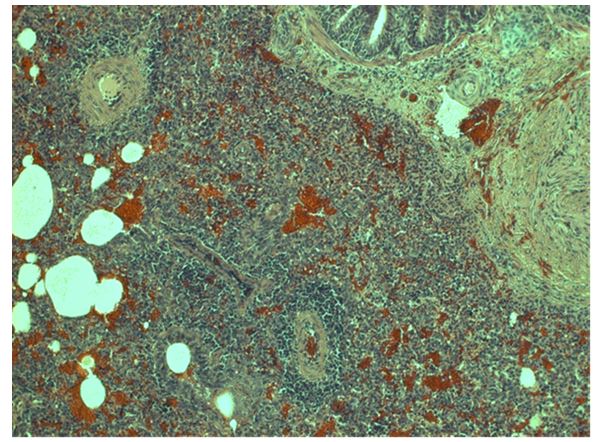
As expected, the lung tissue in the majority of infected animals (23 out of 28) revealed the typical picture of usual interstitial pneumo- nia (UIP). In UIP cases, the originally thin interalveolar septi in the lung became thickened, both due to proliferation of connective tissue and because of the accumulated mononuclear cell infiltratate. In addition, the alveolar capillaries became widened along with occasional focal bleeding, possibly occuring due endothelium cell injury (Figures 2B and 2C). At high power view, there could be recognised that the interstitial infiltrate consists mainly of lymphocytes (Figure 2D). Occasionally (for example in piglet no. 40), the extensive inflammatory infiltration has profoundly altered the original lung structure (Figure 3).
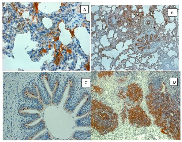
Staining with anti-N antibody showed the presence of PRRSV antigen predominantly in the columnar ciliary epithelium lining the bronchial tree (Figure 4A). At examining such areas in detail, we demonstrated presence of viral antigen in the cytoplasm of epi- thelium cells of the small acinary mucous glands located below the bronchial ciliary epithelium lining, thus in the wall of bronchial tree formed by connective tissue (Figure 4B). Flat cells of the alveolar lining were rarely positive, though their type II alveolar cells could occasionally harbor the N-protein. Howthever, this antigen was mainly seen in macrophages moving from the alveolar space across the thickened interalveolar septa in the direction of local lymphatic capillaries and/or to the sinuses of regional lymph nodes (Figure 4C), where virus was finally deposited. Nevertheless, in some piglets the local lymph nodes did not arrest the virus spread, which namely later on might reach the spleen and/or liver via blood stream. In the spleen therefore, the reticulum cells of sinuses were found positive as well; finally, the N-antigen could be occasionally seen also in the lymphatic follicles (Figure 4D).
Outside of the lung tissue, the N-protein of PRRSV was found in not-hornified squamous epithelium at the pharyngeal area including that covering the tonsils (Figure 5A). Here the virus antigen has occupied deeper layers of the stratified epithelium, namely the still multiplying parabasal cells not excluding those situated in its medium layer. Finally, the virus was also found in the salivary glands. As seen on Fig.5B, the submandibular gland acinar cells could harbor the traced antigen in their cytoplasm. Nevertheless, while the tonsilar and/or pharyngeal epithelium were relatively frequently involved, in salivary gland acinar cells the virus antigen was present relatively rarely. Noteworthy, the real incidence of given antigen in salivary glands was difficult to asses, since such tissue has been met just by chance in the sections examined.
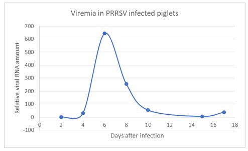
In comparison with lung lesions by infected animals, the development of antibody response in their serum samples was assessed by enzyme immunoassay (i.e. ELISA as documented on Table 2). The given data show that virus-specific antibodies were relative- ly rarely detected on day 11 post-infection, but were regularly found on day 18. Such timing may not be unexpected and can be explained by the dvelopment viremia (Figure 6). This has peaked on day 6 post-infection, being absent by day 10. Interestingly enough, on next day (i.e. on day 11), no serum antibodies were detected, probably due to their abundant interaction with the virus particles. However, the specific antibodies were clearly detected at later intervals (i.e. by day 18), when their levels in serum have increased due to continued intensive production.
The PRR syndrome in piglets is characterized with high mortality, reproductive failure (late-term abortions and stillbirths, pre- mature farrowing, mummified pigs in pregnant sows) and a severe respiratory disease (interstitial pneumonia). The disease occuring in the nursery and among growing/finishing piglets causes significant economic losses to the swine industry worldwide. The corresponding virus (PRRSV) replicates mainly in the porcine alveolar macrophages (PAMs) and dendritic cells (DCs) [21]. The virus also causes persistent infection eliciting antibody dependent enhancement (ADE) and ocassional immunosuppression. Being a member of the family Arteriviridae, it belongs to the order Nidovirales together with the Coronaviridae and Roniviridae families [22]. PRRSV was originally divided into European type 1 and North American type 2 genotypes. Later on, the East Eu- ropean PRRSV isolates have been found to be of the European genotype, but forming different subtypes. A novel virus, namely the Belarusian strain Lena, has been recently characterized as a highly pathogenic East European subtype 3, which differs from European subtype 1 Lelystad and North American US5 strains at genetic as well as antigenic levels [23].
Numerous results suggest that PRRSV may utilize multiple strategies of replication and spread in the infected pigs, including sub- version of the host innate immune response, inducing an anti-apoptotic and anti-inflammatory state as well as developing ADE. The PRRSV induced immunosuppression might mediate apoptosis of infected cells, which causes depletion of immune cells and induces an anti-inflammatory cytokine response due to which the host is unable to eradicate the primary infection. The initial antibodies do not confer protection and can even be harmful by mediating an antibody-dependent enhancement (ADE), since they can facilitate the virus entry of into targets cells in vitro. To characterize the humoral immune response direct enzyme-linked immunosorbent assays (ELISA) can be used including different mainly recombinant PRRSV antigens. For example, the kinetics of antibody responses directed against nonstructural virus coded proteins (nsp) can be analysed in pigs experimentally exposed to the virus [24]. In such case, high antibody reactivities especially against nsp1, nsp2, and nsp7 were noted. Among the latter, nsp7 recombinant protein based ELISA showed good sensitivity and specificity most suitable for diagnostic development especially for identification and differentiation of type 1 and type 2 PRRSV. Several non-structural proteins (such as nsp1, nsp2, nsp5, nsp7 nsp9, nsp10 and nsp11) have been implicated in the induction of IFN-γ and also in the development of the cell-mediated immune response [25]. On other hand, the induction of neutralizing antibodies (NAs) may be delayed and/or their levels may remain low, which is not only the problem of early diagnostic, but is also of importance regarding effective virus elimination. NAs may protect against disease if present in sufficient quantities before infection, but they do not seem to be essential for clearing virus in blood during the course of the infection. PRRSV is able to modulate innate responses, probably through the regulation of IFN-α and IL-10 responses [26].
As described, PRRSV replicates predominantly in the lung alveolar macrophages, can induce prolonged viremia, and cause per- sistent infections lasting for months after initial infection. PRRSV strongly modulates the host’s immune response and changes its gene expression. Studies showed that PRRSV inhibits type I interferons (IFN-β). Regarding cell-mediated responses, development of PRRSV-specific gamma interferon-secreting cells (IFNgamma-SC) and interleukin 4-secreting cells (IL4-SC) in PBMC was examined by ELISPOT assay. Using this technic, no IFNgamma-SC was detected until day 14 p.i., whereas for IL4-SC, such differ- ences were not seen. Concurrently with the onset of viremia and the development of clinical signs, serum haptoglobin levels and interleukin 10 (IL10) in PRRSV-stimulated PBMC-culture supernatants increased significantly. These results are compatible with the model of pathogenesis in which the immune response does not fully control the outcome of infection [27].
The PRRSV replication and its spread in the body subverts the host innate immune response as well when highjacking its lipid me- tabolism and inducing an anti-apoptotic and anti-inflammatory state. The latter is indicated by suppressing the expression of ser- ine proteinase inhibitor 2 (SPI 2), IFN-α, and down-regulation of the expression of pro-apoptotic genes such as B-cell lymphoma 2 (BCL-2) antagonist/killer (BAK) and the BCL-2 associated X (BAX). Whereas BAX resides predominantly in the cytosol, BAK is constitutively localized to the outer mitochondrial membrane; both form toxic mitochondrial pores in response to cellular stress. Furthermore, the APR-1, i.e. the Adenomatous polyposis coli (APC) protein which is a Wnt signaling component along with a microtubule-associated protein SARP3 (several ankyrin repeat protein 3), may be down-regulated. Both were shown to interact with all isoforms of PP1 (protein phosphatase 1). Infections of N-PRRSV viruses resulted in fever and inflammatory response, as indicated by high expression of proinflammatory cytokines and chemokines, adhesion molecules, inflammatory enzymes and their receptors, such as IL-1β, IL8, SELL, ICAM, CCL2, CXCL9, CXCL10, B2M, proteasomes and cathepsins. This was com- pounded by cell death and elevated expression of NFKBIA, XAF1, GADD45A, perforin, granzymes, and cytochrome C, coupled with increased ROS-mediated oxidative stress, as indicated by up-regulated expression of cytochrome b245. Taken together, the N-PRRSV infection may have resulted in an excessive immune and/or inflammatory response that contributed to tissue damage [28-29].