Prosthetic Rehabilitation of Cleft Lip and Palate with Andrew’s Bridge: Case Report
The Cleft Lip and Palate (CLP) is difference of a type of clefting congenital anomaly caused by abnormal facial development during pregnancy. The etiology of CLP is still unknown but there are possible causes such as malnutrition and irradiation during pregnancy, psychological stress, teratogenic agents, infectious agents (viruses), and inheritance. Oro-nasal fistula is the most common complication after the surgical closure of the cleft palate. Prosthetic rehabitation of the anterior maxilla is important for these patients. Retention is the significant factor in the successful prosthodontic rehabilitation of cleft palate patients.
CLP is needing complex treatment strategies scheduled in many years of duration. These patients require various treatments involving a multidisciplinary team, which may include a maxillofacial surgeon, an orthodontist, prosthodontist, an ear nose and throat specialist (ENT), speech therapist and all those professionals who can help provide functional and aesthetic improvement.
Keywords: Cleft Lift and Palate; Prosthetic Rehabiliton; Andrew’s Bridge
The palatal defects can be either congenital or acquired. In Turkey, the incidence of cleft lip and palate is 1 in 1000 live births and majority of these patients are not treated surgically at the appropriate time [1].
Palatal fistula is a common complication arising out of cleft palate repair and its occurence ranges from 0-34 % as observed in various studies [2]. It was found statistically that the width of 15 mm or more had a significant risk of fistula formation.
Smith, et al. classified the palatal fistula into seven types [3].
Type I - referred to bifid uvula.
Type II - means fistula in the soft palate.
Type III - means fistula at junction of the soft and hard palates.
Type IV - means fistula in the hard palate.
Type V- indicates that the fistula at junction of the primary and secondary palates.
Type VI - means lingual alveolar fistula.
Type VII - means labial alveolar fistula.
The patient with a unilateral or bilateral cleft palate, missing anterior teeth and a deficient alveolar ridge presents the problem of restoring the missing teeth and the alveolar ridge [4].
The increasing demand for esthetics in restorations can be met with any of ceramic restoration systems currently available. However, the esthetic value of a cosmetic restoration may be compromised by other factors contributing to the composition of a pleasant smile, such as amount of gingival display, gingival architecture, clinical crown dimensions and tooth position [5].
The major prosthetic treatment to close the soft tissue defects were; obturator, removable flange prosthesis, Andrews bridge and surgical bone augmentation. The fixed partial denture-removable partial denture system was introduced by Dr James Andrews of Amite, Louisiana, USA in 1965, when fixed or removable partial dentures were not successful in treating ridge defects [6]. This system incorporates a fixed component on the abutment teeth with removable pontics. The fixed component is made of porcelain fused to metal crowns that are joined together by casted bar cemented on to the prepared abutments. The removable component consists of acrylic teeth on acrylic base to which metal or plastic sleeve tract are embedded. This technique has the advantage of flexibility in arranging the removable partial denture teeth with minimum extension along with better retention and stability [7]. The indications for fixed-removable Andrews’s bridge system are;
▶ Several missing teeth along with defect in the edentulous ridge;
▶ Failure of removable partial denture because of discomfort related to its palatal extension;
▶ long edentulous space where fixed partial denture has failed;
▶ Cleft palate patients [7-9].
The aim of the present a case having ridge defect, which was restored successfully by using fixed-removable Andrews’s bridge system.
An 18 year old male was referred to the Department of Prosthodontics from the Department of Oral and Maxillofacial Surgery, Ondokuz Mayıs University, Dentistry Faculty, for the prosthetic rehabilitation of the palatal fistula after the primary surgical closure of cleft palate.
Intraoral examination revealed maxilla with a Veau’s class III defect (unilateral cleft lip, alveolus, hard and soft palate) and missing left lateral incisor.
Extra oral examination revealed a surgical scar on the upper lip due to previous surgical correction of cleft lip. Her speech was not intelligible with a nasal twang in her voice lacking resonance.
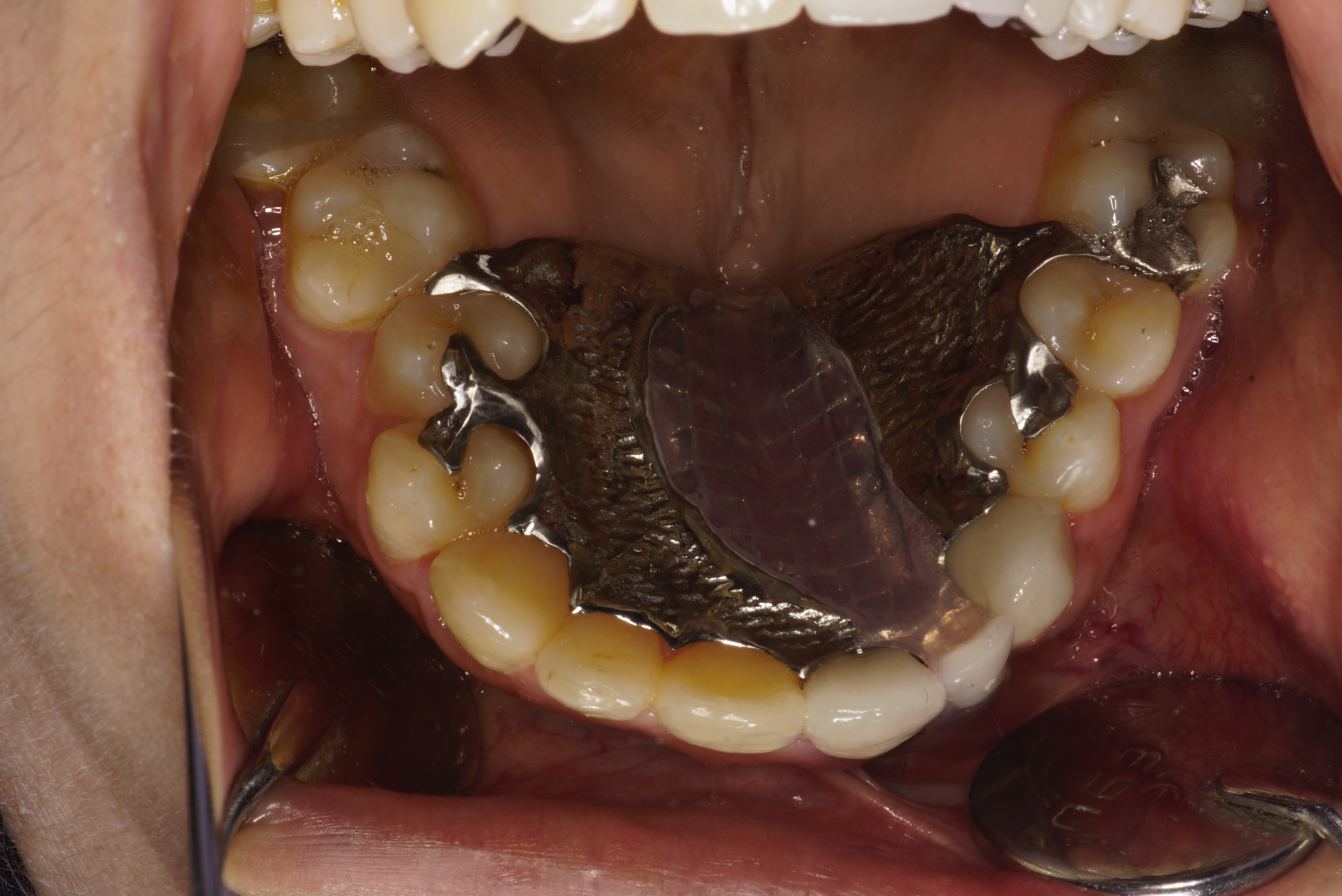
Considering her age and oral conditions we planned to habilitate with an obturator which would be simple in design, light weight, fixed type but easily maintainable prosthesis obturator attached to Andrews’s bridge (fixed removable). The Andrews bridge consists of a metal bar and sleeve. The principle of the system is similar to that of an intracoronal attachment. The pontic portion of the fixed removable partial denture is removable. This permits access to the underlying tissue for cleansing purposes. The Hader bar was planned between 21 and 23 attached to metal ceramic crowns
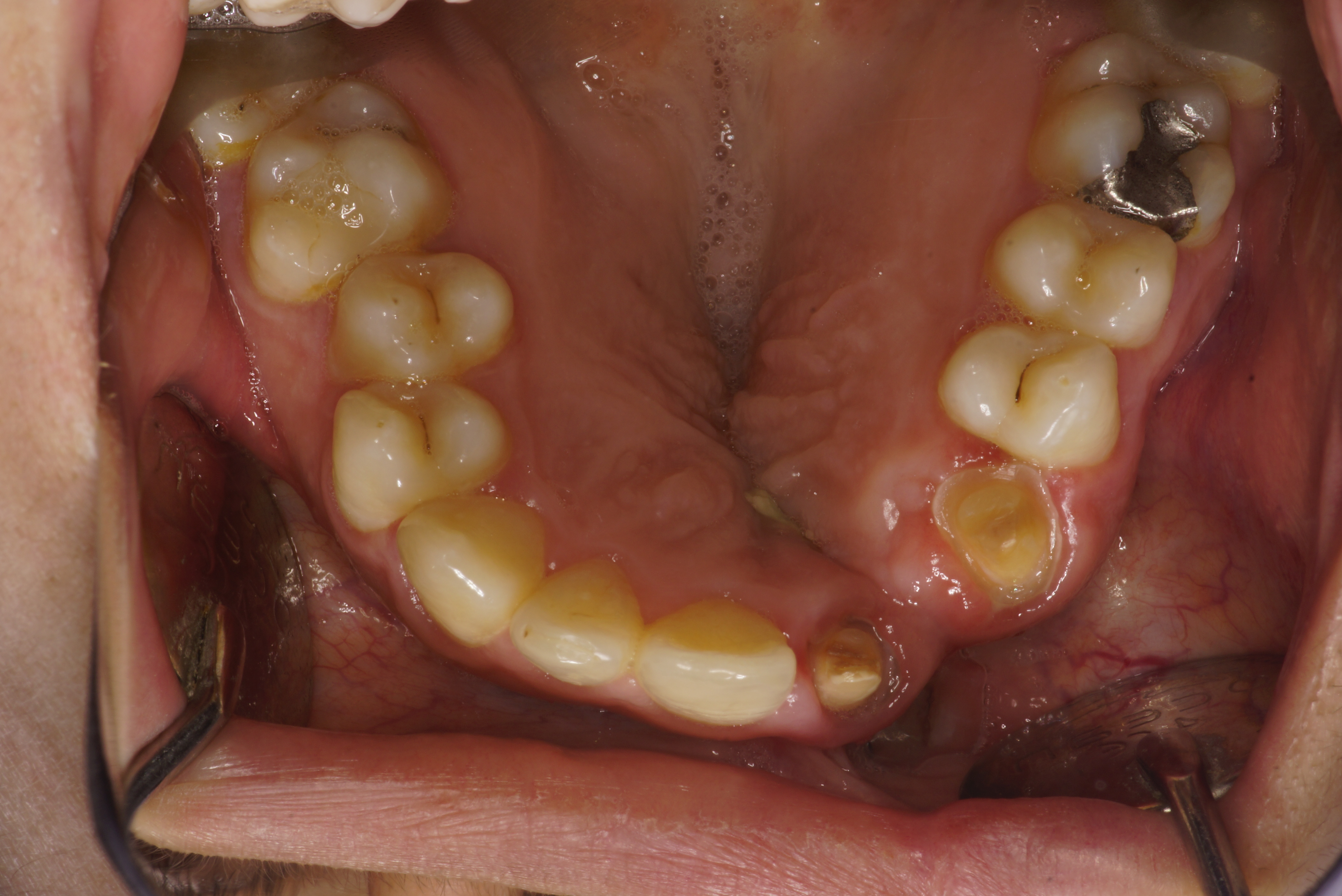
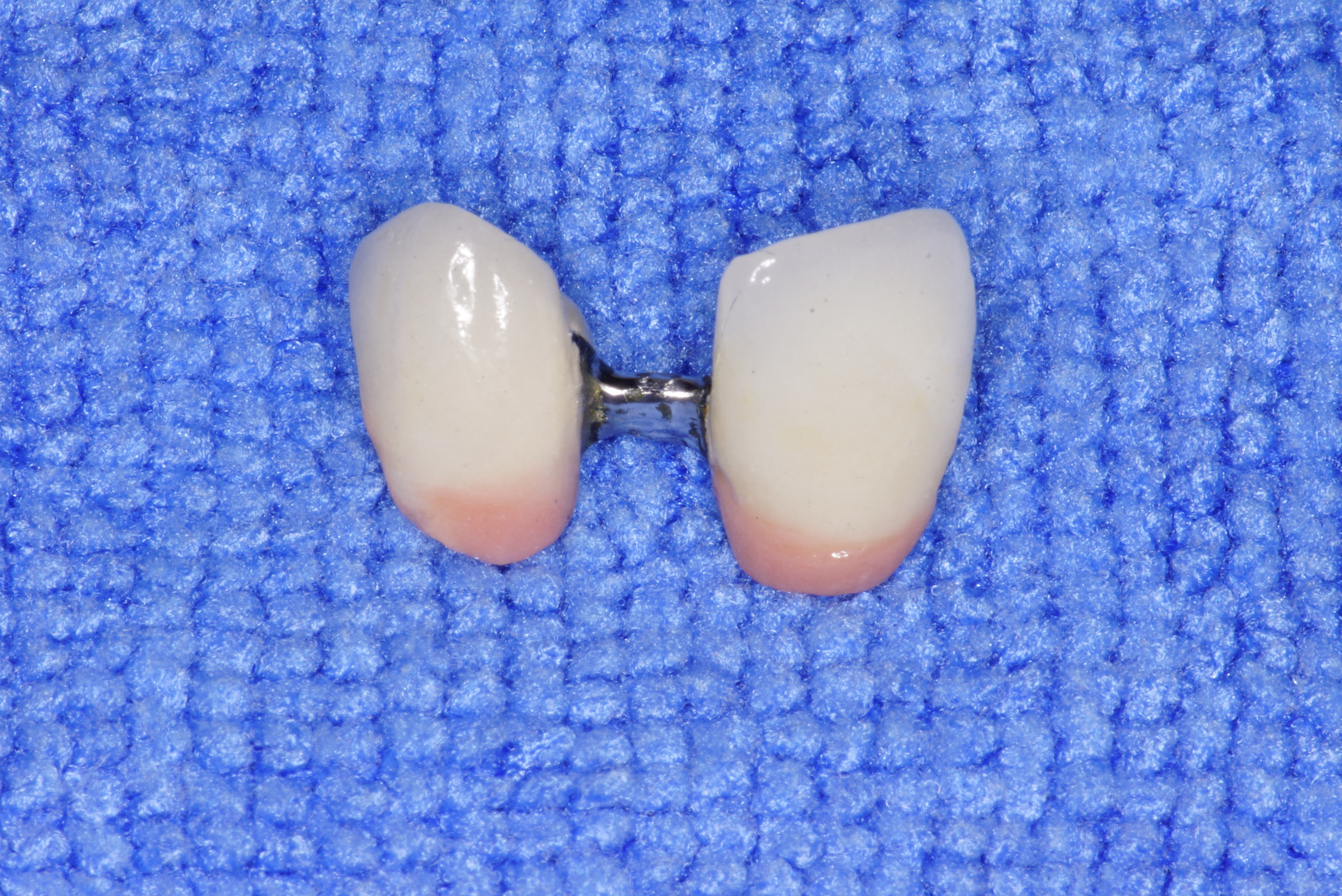
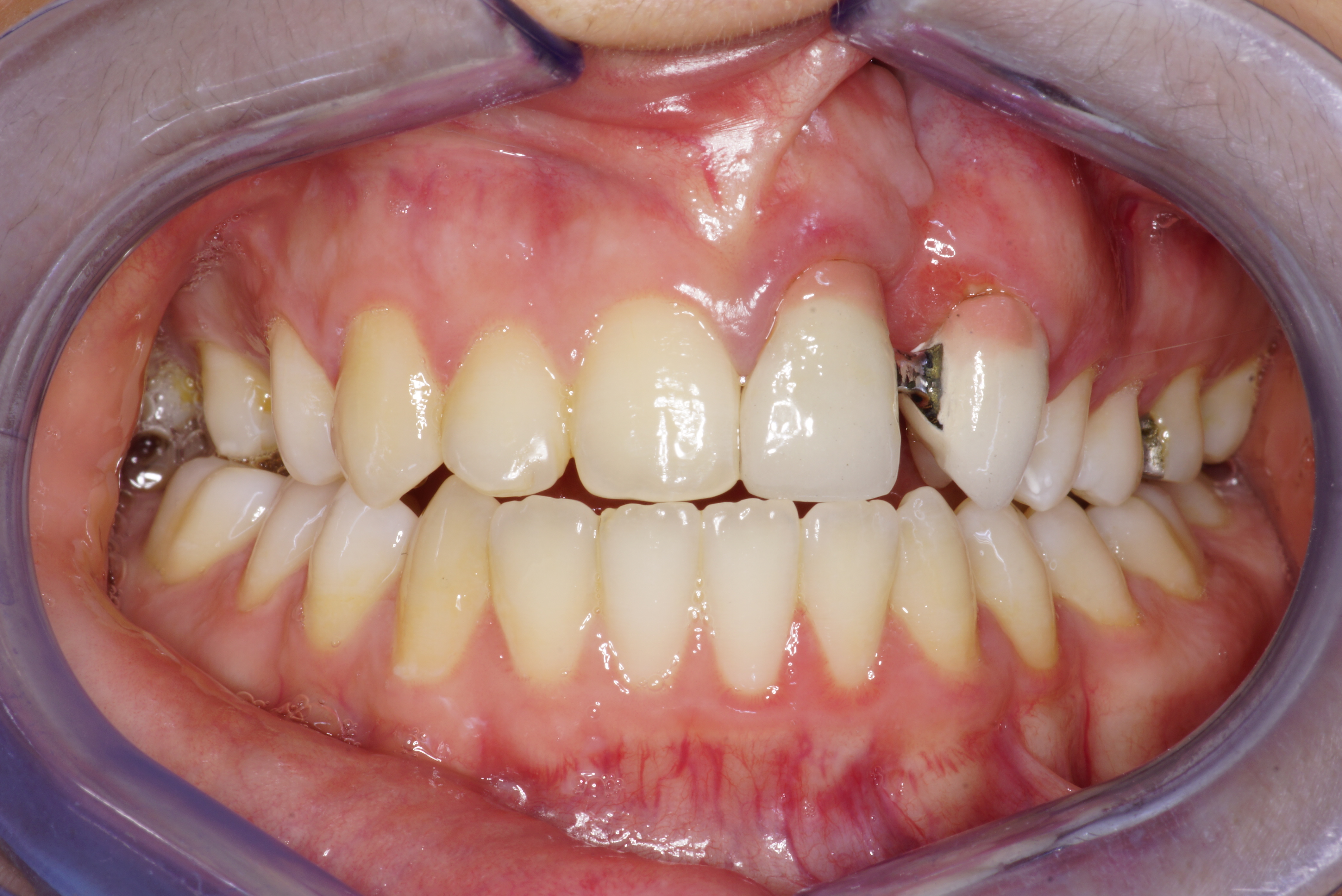
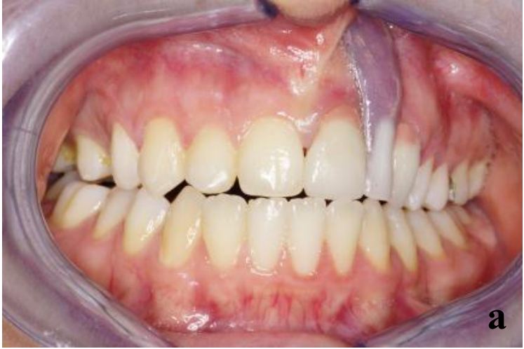
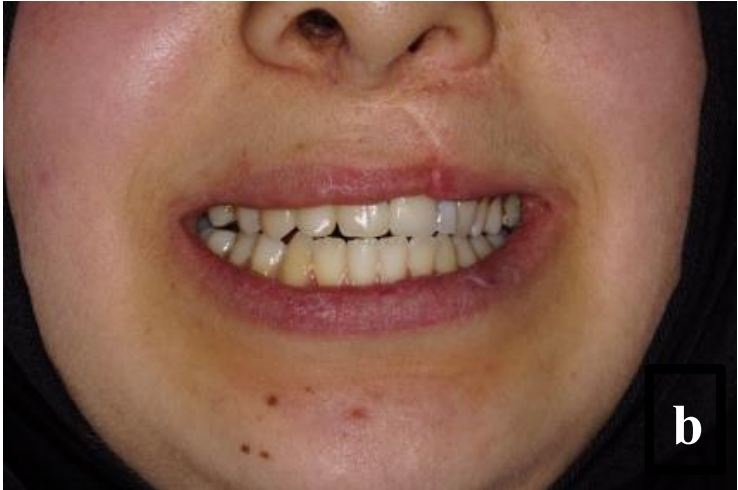
The selected abutment teeth were prepared for receiving metal ceramic crowns (Figure 1). Impressions were made using the putty wash impression technique(Express STD Putty 3M, ESPE, USA and Express Light body STD Quick Wash, 3M ESPE, USA) and master casts were poured in the dental stone (Type IV die stone- Gyproc, InZhermack, Rovigo, Vêneto, Italy). Wax patterns were fabricated on the prepared abutment teeth and were connected using a preformed plastic bar attachment. This entire assembly was then cast in chrome cobalt alloy. The finished and polished metal framework was tried in the patient’s mouth for proper fit and clearance between the bar attachment and underlying soft tissues (Figure 2 and 3). The missing teeth were arranged on the wax occlusal rim fabricated on to the edentulous area of the cast and tried for aesthetic approval by the patient. The removable part of Andrew’s bridge was then fabricated using heat cured polymethylmethacrylate (PMMA) resin (Dental Products of India DPI, Mumbai). Plastic clip and metal housing were placed on to the cast bar before packing of acrylic resin. Metal copings were then veneered with ceramic and the whole restoration was finished and polished (Figure 4). The fixed component of the Andrew’s system was cemented over the prepared teeth. After 1 hour a removable component was inserted and occlusal adjustments were carried out (Figure 5aand 5b). The patient was recalled after the first day then after a week. The patient was happy with the aesthetics and functions of the prosthesis and was advised for regular recall visits.
The most commonly seen defects are the combined Class III defects (56% of cases), followed by horizontal defects Class I (33 % of the cases) [10]. Vertical defects were reported to be found in 3% of the patients [11]. Large vertical and horizontal bone defects pose a prosthodontic challenge as it is difficult to restore esthetics and function along with the complete closure of the defect. Such clinical conditions are not successfully treated by conventional fixed or removable prosthesis.
Prosthodontist play a very important role in complete rehabilitation of cleft lip and palate patients for successful rehabilitation of such patients, a team approach of concerned specialist with phase wise, unanimous and systematic treatment plan is essential. Prosthodontic rehabilitation of these patients is usually around the age of 20, before which the patient will be treated by surgeons for surgical primary closure, orthodontists for alignment of teeth and finally by prosthodontist for the replacement of missing teeth and obturation of the defect.
Palatal fistula is a common complication of cleft palate surgical repair observed in different studies and to correct inevitable sequel of the surgery, the prosthetic rehabilitation with obturator is an auxiliary or complementary treatment to surgical treatments.
The advantages of the Andrew’s bridge system are adequately reported in the literature, which includes better aesthetics, hygiene along with better adaptability and phonetics. It is comfortable and economical for patients. There is no palatal extension as in the case of removable partial dentures. Good soft tissue response due to less soft tissue impingement. This type of prosthesis is more retentive and stable with minimal extension. The system avoids transfer of unwanted leverage forces to the abutment teeth by acting as a stress breaker [6,7].
Andrews Bridge System is a fixedremovable prosthesis that is indicated in patients with large ridge defects. The functionally fixed prosthesis successfully replaces the missing teeth along with complete closure of the defect, restores speech and esthetics.