Rhinolithiasis: A Case Report and Review of the Literature
Background:Rhinolithiasis is a rare clinical entity usually resulting from the accumulation of retained foreign bodies in the nasal cavity.
Aim: The aim of this case report is to emphasize the importance of this clinical entity, the diagnostic difficulties that it presents by describing an unusual case of a misdiagnosed rhinolith that have been evolving for 7 years with no major local complications
Case Report: We report the case of a 32 years old patient presenting a 7 years right nasal obstruction and fetid rhinorrhea misdiagnosed and treated as sinusitis, revealing after endoscopic and radiologic exploration, a giant right rhinolith.
An endoscopic extraction under general anesthesia was performed.
Results: A clinical evaluation after 3 months showed the regression of the symptoms with no sign of nasal obstruction.
Conclusion: Unexplained, persistent unilateral nasal symptoms should lead the clinician to look for a rhinolith in order to avoid misdiagnosis and local complications.
Keywords:Nasal Obstruction; Giant Rhinolith; Endoscopy
Rhinolithiasis is a rare condition often neglected or unknown, and corresponds to a solid calcification by gradual deposition of calcareous salts around a central resorbable or non-resorbable foundation of varying shape and size [1].
Though infrequently observed, rhinolith can be the source of foul smell from the nose and therefore a social concern for the patient [2].
Endoscopic examination can confirm the diagnosis which can be supported with a CT scan. Complete resolution of symptoms occurs after surgical removal. Early diagnosis can avoid potentially serious complications related to the chronicization of the resultant irritation, with a real risk of superinfection. The purpose of this case report is to emphasize the importance of this clinical entity and the diagnostic difficulties that it presents, by describing an unusual case of a misdiagnosed rhinolith that have been evolving for 7 years with no local major complications.
Although this entity tends to disappear in developed countries, it’s still influenced by different factors in developing countries such as difficult access to healthcare, patient noncompliance and the impact of socio economic conditions.
A 32 years old construction worker with no particular background consulted to our department for 7 years’ anterior and posterior fetid intermittent rhinorrhea, right nasal obstruction and chronic headache.
The patient consulted different general practitioners. They concluded several times that it was an acute sinusitis and antibiotics with nasal irrigation were prescribed.
Due to the intermittency of symptoms and the patient noncompliance, no correct follow-up nor proper endoscopic examination by an ENT specialist have been done.
After years of self-medication and long term persistence of his clinical symptoms, the patient decided to consult in our department.
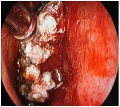
The endoscopic examination, after aspiration of the purulent secretions, revealed in his right nasal cavity an irregular dark gray mass with a firm hard consistency and a deviated septum to the left.
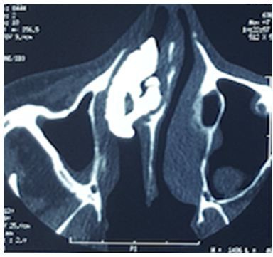
A CT scan of paranasal sinus revealed several masses with a calcific density in the right nasal cavity responsible of a septum deviation to the left with no sign of osteolysis (Figure 1).
The diagnosis was assessed through endoscopic and radiological findings.
An endoscopic extraction under general anesthesia was performed after fragmentation of the giant rhinolith (Figure 2).
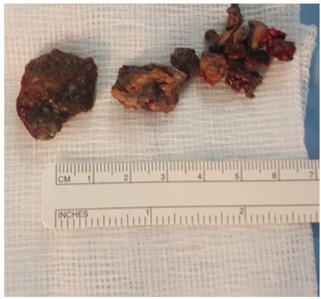
The per operative exploration noted several stones presented in a giant rhinolith occupying the whole nasal cavity.
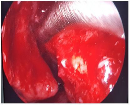
The endoscopic verification showed the complete emptiness of the right nasal cavity (Figure 3).
Histopathology examination revealed fragments of non-viable tissue with areas of calcification suggestive of rhinolith.
A clinical evaluation after 3 months showed the regression of the symptoms with no sign of nasal obstruction.
The first case report of a rhinolith in the nasal cavity was published in 1654 by Bartholini who described a nasal hard foreign body that had grown around a cherry stone, and the first chemical analysis was conducted by Axmann in 1829 [2].
Rhinoliths can be exogenous and endogenous calcified nasal masses depending on whether or not a nucleus, around which the incrustation has been deposited, can be found.
The ones that have developed around nonhuman material introduced into the nose such as stones, forgotten nasal swabs, or similar objects are termed exogenous however rhinoliths that have developed around the body’s own material such as ectopic teeth in the maxillary sinus, bone sequesters, dried blood clots in the nasal cavity are known as endogenous [3,4].
The pathogenesis of this entity remains unclear, the most probable theory is the deposit of calcium magnesium and other organic substances due to the local inflammation caused by the of presence foreign nasal mass.
Rhinoliths are often asymptomatic or with only subtle signs therefore misdiagnosis is frequent. Our patient symptoms were misdiagnosed for 7 years and wrongly treated as acute sinusitis with antibiotics and nasal irrigation.
The main symptoms are usually chronic fetid discharge, nasal obstruction, cacosmia, epistaxis and headache [5]. If undetected for many years rhinoliths may grow large enough to cause nasal obstruction and distortion or destruction of surrounding structures.
Khairul Bariah, et al. reported a case of a giant rhinolith complicated with dystrophy of nasal structures and dehiscence of the lamina papyracea of the right side [6]. Kharoubi reported an unusual case of bilateral rhinolithiasis subsequent to destruction of the posterior nasal septum [7].
In 2010, he also reported 700 cases of nasal foreign bodies with one case of nasal perforation, 5 cases of epistaxis and 10 cases of local infection [8].
In our case, even though the duration of development of the rhinolith was significant, our patient only presented a septum deviation with no other local major complication.
A wide variety of pathologic findings present with unilateral sinonasal symptoms: chronic rhinosinusitis, benign tumors (antrochoanal polyp, nasal polyposis, concha bullosa mucocele), malignant tumors. Therefore, a careful history examination and radiographic studies can often determine the responsible disease process.
The examination should include anterior rhinoscopy and rigid endoscopy [9].
Endoscopy also allows a lesional assessment: septal perforation, synechia, granuloma and reaction polyp.
Computed tomography of the paranasal sinuses can accurately support the diagnosis through the presence of calcified masses in the nasal cavity, it can also determine the site and size of the rhinolith and identify any coexisting sinus disease which may require treatment [10].
Its findings are very useful in the choice of the process therapeutic approach (approach, prediction on the difficulties of extraction) and for differential diagnosis that include osteoma, calcified polyps, bone sequestration (syphilis, radiotherapy), osteosarcoma and chondrosarcoma [7].
Treatment of choice is endoscopic surgical removal. External approaches may be necessary in rare cases of giant rhinoliths [11].
Complications and coexisting diseases should also be an important step in the therapy approach in order to guarantee satisfying results.
Unexplained, persistent unilateral nasal symptoms should lead the clinician to look for a rhinolith in order to avoid misdiagnosis and local complications.
Rigid nasal endoscopy and CT imaging are very useful especially for diagnosing hidden rhinoliths and in showing their extension.
Endoscopic surgical removal remains the treatment of choice.