Solitary Gastric Metastasis of Breast İnvasive Lobular Carcinoma
A 52-year-old female patient with solitary gastric metastasis from breast invasive lobular cancer without any other organ involvement is presented in this study. Solitary gastric metastasis of breast cancers is very rare. It can be diagnosed incidentally by endoscopic biopsy and immuno-histo-chemical analysis. Symptoms and clinical findings are not different from primary gastric cancers. Prognosis of solitary gastric metastasis is good after surgical treatment according to patients with systemic metastasis.
Keywords: Breast; Invasive Lobular Carcinoma; Metastatic Gastric Cancer
In parallel with the increases in the lifetimes of individuals and the rates of chronic diseases in the 21st century, cardiovascular diseases are in first place in mortality statistics, while cancers are in second place. Cancer is one of the major causes of death globally, accounting for nearly 10 million deaths in 2020. The most common cancers were reported as breast, lung and colorectal cancers [1]. Lung, prostate, colorectal, gastric and liver cancers are the most common types of cancer in men, while breast, colorectal, lung, cervical and thyroid cancers are the most common among women [2,3].
After a certain time, cancers spread regionally and systemically by metastasizing. While metastasis to the stomach is not very frequently encountered, it can only be seen in breast cancer and sometimes renal-cell carcinoma [4-7]. In addition to bone, lung, liver and brain metastases, gynecologic organ and peritoneum involvement can be seen in metastasized and advanced-stage breast cancer cases [8-10]. Approximately 90% of breast cancers are invasive ductal cancers, whereas 19% are invasive lobular cancers. Breast cancers that metastasize to the stomach are usually invasive lobular cancers (ILC).
Breast cancer metastasis in the stomach is mostly accompanied by systemic metastasis, and its incidence is lower than 0.1% [10,11]. Gastric metastasis alone, without the involvement of other organs or lymphatic metastasis, is much rarer. In this study, a breast cancer case with solitary gastric metastasis, which is very rare, is presented and discussed along with information in the literature.
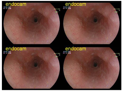
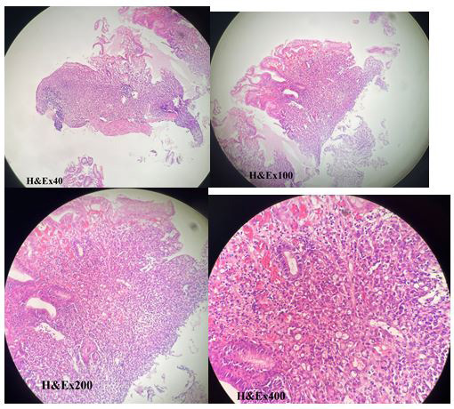
We report a case of gastric metastasis from ILC of the breast in a 52-year-old woman who had progressive epigastric discomfort for 4-5 months. The initial diagnosis was gastric signet-ring cell carcinoma based on endoscopic findings and gastric biopsy findings. In the examination of the gastric biopsy material obtained after detecting superficial ulcerative lesions in the gastric cardia and fundus and thickening of mucosal folds in upper gastrointestinal endoscopy (Figure 1), a malignant tumoral infiltration involving the entire mucosa in the corpus region and containing superficial ulceration was observed. The tumor cells did not show a specific histological differentiation. In many loci, there were intracytoplasmic mucin-like inclusions in the tumor. The histomorphological findings in the lesion initially indicated a signet-ring cell cancer. In the immunohistochemical analysis (IHCA) conducted for a definitive diagnosis, E-cadherin staining was negative, C-erbB2 was negative, CK20 and CDX2 were negative, CK7 and GCDFP-15 were positive, and there was strong nuclear staining (98% +) with estrogen receptors (ER), as well as heterogeneous moderate nuclear staining (5-10% +) with progesterone receptors (PR). Diffuse strong cytoplasmic positive staining was observed with cytokeratin-19 and mammaglobin. It was determined that E-cadherin was negative and showed diffuse expression loss, while diffuse strong nuclear positive staining was seen with GATA-3. In the assessment with these immunohistochemical data and morphological findings, it was considered that the case could be metastasized breast cancer (Figure 2).
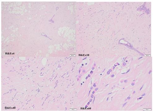
There was no known history of breast cancer in the patient, but she stated that she had been in periodic follow-up for 3 years for a benign lesion in the BIRADS-3 category in her left breast. To reveal primary tumor localization, bilateral breast MRI and whole-body positron emission tomography (PET/CT) were performed on the patient. There was no pathological appearance suggesting malignancy in the MR images. However, PET/CT showed moderate 18-fluorodeoxyglucose (FDG) uptake in the region of the lesion that was previously assessed as benign in the left breast (SUVmax: 2.7). There was no pathological FDG uptake in any organ or region in the body except for the stomach. Trucut biopsy was taken from the region identified with PET/CT. In the microscopic examination, there were ducts showing cystic dilatations and epithelial cells with hyperchromatic nuclei and eosinophilic cytoplasm containing proliferated lobules, and the findings were compatible with ILC. As a result, invasive lobular carcinoma of the breast was diagnosed by core needle biopsy (Figure 3).
In IHCA, ER, PR, CK7 and GCDFP-15 were positive, and CK20 was negative. With these findings, for the diagnosis of solitary gastric metastasis of breast cancer, curative surgical treatment was planned for the breast and the stomach. In the same surgical session, modified radical mastectomy (MRM) + axillary lymph node dissection and total gastrectomy + D2 lymph node dissection were performed. In the immunohistochemical examination, similar to the case in the breast biopsy, the tumor cells had diffuse nuclear positive staining with GATA-3, and they had expression loss with E-cadherin. In the tumor cells, C-erbB2 (clone 4B5) was negative, and there was 100% nuclear staining with ER. With histopathological evaluation and these IHCA results, it was concluded that there was metastatic ILC in the stomach (Figure 4). As the tumor cells were 100% ER-positive and chemotherapy-resistant, docetaxel (DTX) + dexamethasone treatment was started for the patient, and she was registered for routine follow-up.
The clinical symptoms of the gastric metastases of breast cancers are not different than other gastric cancers. Patients may display non-specific symptoms such as dysphagia, dyspepsia, loss of appetite, early satiety, nausea, vomiting and upper gastrointestinal hemorrhage [4,11]. As the tumor is usually diffusely metastasized in the submucosal plane in gastric metastases, there might not be noticeable radiological or endoscopic findings in patients in the early period, and cases are frequently identified incidentally as a result of biopsy [10,12,13]. Typically, gastric metastases are confined to the submucosa and muscularis, and thus, mucosal biopsy specimens might be false-negative. When gastric cancer is diagnosed in a woman with a history of breast cancer, breast cancer metastasis may come to mind, but it is not possible to predict this in other patients. In the case reported in this study, in the upper gastrointestinal endoscopic examination of the 52-year-old female patient carried out due to dyspeptic complaints, cancer was diagnosed by the histopathological examination of the biopsy material taken from hypertrophic gastric mucosal folds and superficial linear ulcers. In advanced-stage breast cancers with systemic metastasis, it is possible to explain gastric metastasis with other metastasis mechanisms, but cases of solitary gastric metastasis, without the involvement of other organs, require clarification. As no tumor invasion was detected in the axillary and perigastric lymph nodes of the reported case, it was presumed that the cancer had hematogenous metastasis through the aberrant venous collateral route.
In the gastric metastases of breast cancers, the microscopic findings usually have similar characteristics to those in signet-ring cell and cohesive carcinoma cases. In these patients, IHCA provides the most important clues for the diagnosis. In IHCA, E-cadherin negativity and ER and PR positivity are significant. While weak ER and PR positivity is seen in a few loci in gastric cancers in rare cases, the absence of staining with E-cadherin is especially typical for breast ILC [12,14,15]. CK20, which is positive in gastric cancers, is negative in breast cancers. In addition to these characteristics, CK and GCDFP-15 positivity and CK20 negativity in IHCA are findings that support the diagnosis of metastatic breast cancer
Breast cancers with gastric metastasis that are reported in the literature are usually advanced-stage ILC patients with systemic metastasis. This is why most patients are not suitable for surgical treatment, and they are treated with chemotherapy and/or hormonotherapy. The life expectancy of patients varies between 9 months and 23 months (11 months on average). In breast cancers with solitary gastric metastasis, the main treatment method is surgery, and the prognosis is better. The life expectancy of these patients is longer than 3 years [10-12]. The case reported in this study was also an ILC case like similar cases in the literature. Nevertheless, it was considered worth reporting as it involved only gastric metastasis and no other organ involvement.
The surgical treatment performed in solitary gastric metastases is not different than treatments performed in primary gastric cancers. In cases where resection is applicable, favorable results can be obtained by radical surgical intervention. Palliative interventions and adjuvant chemoradiotherapy/hormonotherapy programs should be provided in patients for whom surgical treatment is not applicable.