Sub-Laminar Wiring in Spinal Fractures – A Viable Alternative
Background: In Sri Lanka toddy tapping is a common form industry in the coast line villages. Falls from palm trees of forty feet is a common occupational hazard. Spinal fractures are common in these patients.
Methods: A retrospective analysis of a personal case series of the two authors was undertaken.
Results: A total of 37 pedicle screw fixations and 58 sub laminar wirings were done during a period of five years from 2004-2009. Follow up time was 6 months to 4 years (mean 3 years).
Of the 58 sub laminar wirings, 41 were for lumbar fractures and 17 for thoracic fractures. Thirty two of the 58 sub laminar wirings were for injury due to falls from scaffoldings. Twenty one were toddy tappers who fell off the coconut palms. Five of the 37 pedicle screw fixations were done for thoracic fractures and 32 were done for lumbar fractures. Fourteen patients had falls from scaffoldings. Thirteen posterior fusions of the cervical spine were done. Seven of these were caused by tree climbing accidents.
Three patients with sub-laminar wiring developed superficial wound infections while two had to have their metal work taken out due to deep infection. One thoracic sub laminar wiring patient had a CSF leak and two patients with lumbar pedicle screw fixation had cerebro-spinal fluid (CSF) leaks. Three patients with pedicle screw fixations to the lumbar spine had metal work failure due to non-union. One patient with pedicle screws had a re-fracture while three patients with sub laminar wiring of the thoracic spine had pneumothoraxes.
Conclusion: Sub laminar wiring is a cheap stable method of fixation for traumatic spines which can be used in relatively under- equipped centers as well.
Keywords: Henoch-SchÖlein purpura nephritis; IgA nephropathy; Oxford Classification; Children; Immune Complex
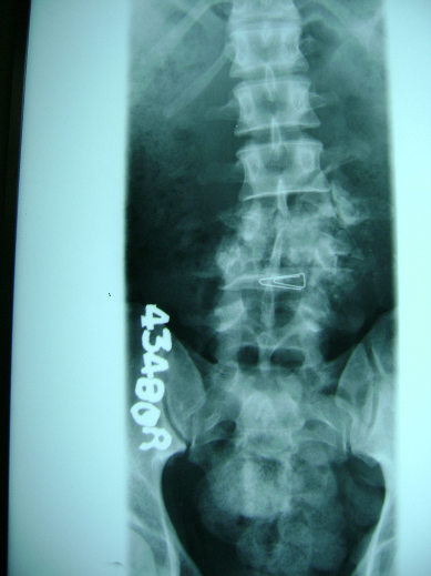
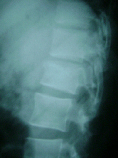
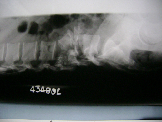
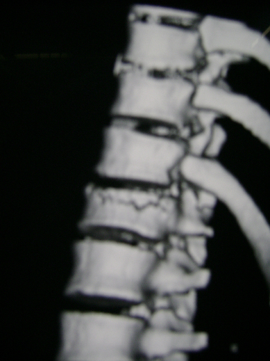
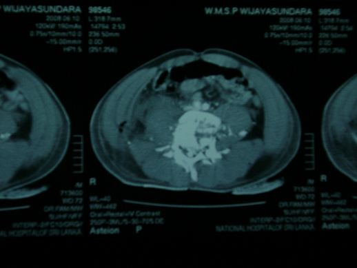
In the coastal regions of the island nation of Sri Lanka (Ceylon) toddy tapping form coconut trees is a common occupation which is part of the vast industry of production of juggery, coconut vinegar, toddy and arrack (a whisky brewed from coconut sap). The harvesting of the sap is done by toddy tappers who climb these nearly forty foot high palm trees and cross from one tree to another using rope bridges. Falls from these rope bridges are not uncommon (Figures 1,a,b and 2,a,b) as rats that are often attracted to the coconut sap take the same root up the tree and damage the rope bridges that are used by the toddy tappers.
Hence falls from these tall trees with resultant spinal fractures is common in the coastal belt of Sri Lanka.
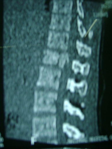
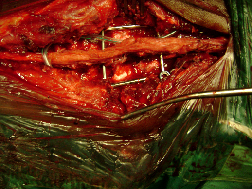
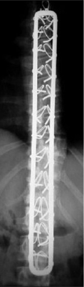
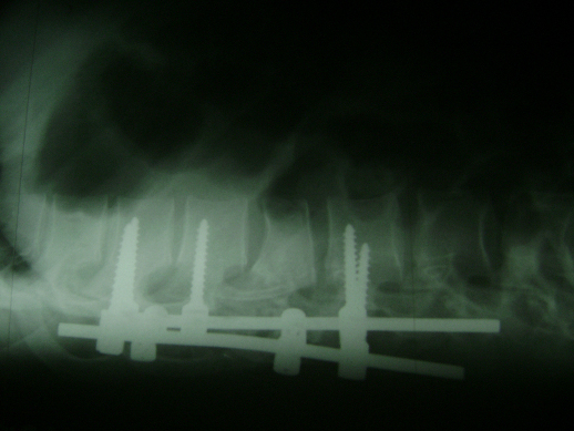
In this retrospective case series, cases treated operatively by the authors were looked upon. A total of 37 pedicle screw fixations (Figure 3) and 58 sub laminar wirings (Figure 4) were done during a period of five years from 2004-2009.
The choice between sub-laminar wiring and pedicle screw fixation depended on the availability of implants in the state health sector at the given instance. Both procedures were undertaken with the patient prone and the spine exposed through the posterior midline approach (Figure 5). Spinal decompression was under taken with laminectomy where indicated and posterior instrumentation was undertaken. Cancellous bone graft from the iliac crest was used for a posterior fusion in both sub-laminar wiring as well as following pedicle screw fixation (Figure 3, 4 and 5). In both the groups two levels above and below were fixed. The criteria for fixation were all unstable fractures based on Denis’s three column classification, with and without neurology. Anterior instrumentation was not under taken. The mean follow up was 3 years with a range of six months to 4 years.
Of the 58 sub laminar wirings, 41 were for lumbar fractures and 17 for thoracic fractures. Thirty two of the 58 sub laminar wirings were for injury due to falls from scaffoldings. Twenty one were toddy tappers who fell off the coconut palms (Table 1).
Five of the 37 pedicle screw fixations were done for thoracic fractures and 32 were done for lumbar fractures. Fourteen patients had falls from scaffoldings (Table 2).
In addition to thoracic and lumbar spine trauma we did encounter cervical spinal injuries which were not included in this series. Considering the causes for spinal fractures in the 95 patients who either had sub-laminar wiring or pedicle screw fixation, the greater majority were due to falls from tree climbing related accidents (Table 3).
We under took surgical fixation of fractures with neurological deficit and fractures which were unstable but, had no neurological deficit. The neurological deficit was assessed using the Frankel Grading system. Frankel grade D and E were fixed when the fracture was unstable. Frankel A, B and C were fixed to allow early mobilization and easy rehabilitation.
Out of the 37 patients who underwent pedicle screw fixation 21 had some neurological deficit while the balance were patients with unstable fractures without neurological deficit.
Forty eight of the 57 patients who underwent sub-laminar wiring had complete or partial neurological deficit while the balance had unstable fractures only.
Three patients with sub-laminar wiring developed superficial wound infections while two had to have their metal work taken out due to deep infection. One thoracic sub laminar wiring patient had a CSF leak and two patients with lumbar pedicle screw fixation had CSF leaks (Table 4).
Three patients with pedicle screw fixations to the lumbar spine had metal work failure due to non-union. One patient with pedicle screws had a re-fracture while three patients with sub laminar wiring of the thoracic spine had pneumothoraxes.
As mentioned above toddy tapping is one of the major causes of falls from heights resulting in spinal trauma in Sri Lanka in this series. Lamawansa and Piyathilaka reported a 1.6% incidence of falls causing injury in a Sri Lankan rural community. However, the study does not look into the causes of falls and the resultant injuries [1].
We treated patients with unstable fractures with neurological deficit complete and partial to stabilize the fracture and to enable early mobilization in the case of patients with complete neurological deficit so that complications of prolonged bed bound nursing were minimized. Patients with no neurology but with unstable fractures also underwent fixation as a precautionary measure to prevent spinal cord damage due to the unstable nature of the fractures and to enable early mobilization.
The choice between the fixator used was dependent on the availability of the relevant implants in the state health sector which is a free health system to all Sri Lankans. Pedicle screw fixation is a more technically demanding form of fixation requiring the use of the image intensifier where-as sub-laminar wiring is less demanding on resources and the necessity of an image intensifier is not required. This has been a favoured technique of spinal fixation in base hospitals and rural district general hospitals where an image intensifier may not be available throughout Sri Lanka.
The down side of sub-laminar wiring is the extensive exposure required and the potential for tears in the dural sac during insertion of the wires. This rather extensive dissection and longer operation times may be partly responsible for the slightly higher infection rates noted in the group of patients who had sub-laminar fixations done.
The nature of the two implants themselves where the sub-laminar wiring system with Hartshill ring fixation is a load sharing device and the pedicle screws being a load bearing device could account for the difference noted in the metal work failure rates seen in the two groups. Posterior on lay bone grafting on the freshened laminae and transverse processes were used as a bony fusion to prevent metal work failure. We noted failure of metal work in the pedicle screw fixation group more than in the sub-laminar wiring group. Non-union of the bone graft and fracture itself could have resulted in the failures noted.
Using the word combinations of “sub-laminar wire fixation in spinal trauma”, “sub-laminar wires in spinal trauma”, “Sub-laminar wires, spinal trauma”; no articles were found on a PUBMED search. However, the Hartshill ring and sub-laminar wires construct has been in use for corrective surgery of spinal deformities.
Kerboul and Courtois described the use of the Hartshill ring and sub-laminar wire construct in the correction of scoliosis and in posterior spinal instability [2]. In a series of 92 patients they encountered 08 cases of device failure.
The use of a Hartshill ring and sub-laminar wires has been documented in the literature for correction of spinal column deformities [3]. In this paper the authors have noted an average of 64% correction rate with the use of this fixatory technique [3].
Pedicle screw fixation has been the favoured technique of fixation in spinal trauma with or without anterior instrumentation and very good success rates have been reported. Metal work failure has been a minimum in most published series.
Xu and Jiang reported no metal work failure in series of 94 patients who underwent pedicle screw fixation in burst fractures of the thoraco-lumbar spine at a mean follow-up of 22 months [4]. Deng, Zhao et al found in their paper on a series of 27 short segment pedicle screw fixations of the thoraco-lumbar spine that it was an effective method of spinal fracture fixation [5].
Sub laminar wiring is a cheap stable method of fixation for traumatic spines which can be used in relatively under- equipped centers as well.