Systemic Hypertension with Ultrasound Findings of Multiple Simple Hepatic Cysts and Simple Left Renal Cyst in an Elderly South Eastern Nigerian Female: A Case Report
Hypertension is a major risk factor of cardiovascular disease, as well as a common chest radiological finding in elderly patients. The present report aimed to determine that link between hypertension and multiple hepatic cysts and simple renal cyst. We report a rare radiological case of systemic hypertension, with ultrasound incidental findings of multiple liver cysts and left upper pole simple renal cyst in a 72 years old woman from south east Nigeria who had history of chronic cough with purulent sputum, chest pain, palpitation and abdominal discomfort or pain. Her BP was 180/110 mmHg, and her heart rate was 100 bpm. Chest radiograph revealed a prominent aortic unfolding or knuckle. ECG revealed sinus tachycardia with no acute ST-T wave changes. Ultrasound studies revealed multiple, rounded thin-walled, anechoic cysts of large size, located within the right and left lobes of the liver. The large cysts on the right lobe were seen pressing slightly on the inferior vena cava. A left simple renal cortical cyst was seen. The cyst appeared to be large in size more than 1 cm in diameter, which is a characteristics of simple renal cyst related to hypertension. No ascites and lymphadenopathy were found in the abdomen. The presented patient was managed using ultrasound guided percutaneous drainage of the hepatic cysts and simple renal cyst to alleviate her symptoms. Ciprofloxacin regimens were administered to her after the procedure against the possibility of infection. Her blood pressure came down to 130/87mmHg post drainage procedure.
Keywords:Hypertension; Hepatic Cyst; Simple Renal Cyst; Chest Radiography; Ultrasound; Nigeria
List of abbreviations:CTR: Cardiothoracic Ratio; BP: Blood pressure; ECG; Electrocardiogram PA: postero-anterior; GE: General Electric; USA: United States of America
Systemic hypertension is high blood pressure in the systemic arteries. These are vessels that carry blood from the heart to the tissues of the body, apart from the lungs. Examples of such vessels are the great vessels; which are superior vena cava, inferior vena cava, pulmonary arteries, pulmonary veins and aorta. Hypertension is mostly prevalent in the elderly and represents a major risk factor for most cardiovascular complications like stroke, cognitive dysfunction and coronary cardiac disease [1-3].
Hepatic cystic lesions constitute an inclusive heterogeneous cluster with regard to pathogenesis, diagnostic findings and clinical presentation [4]. Simple hepatic cysts are the most common cystic diseases which are increasingly found by chance or coincidentally on abdominal imaging procedures, such as ultrasound, computed tomography and magnetic resonance imaging (MRI) [4]. The widespread of simple hepatic cysts in the human population varies from 2-18%. While they are usually asymptomatic, large hepatic cysts may become symptomatic; which may include infection and rupture into the peritoneum [5].
Simple renal cysts are the most common cystic deformation found in adults [6]. They are mostly common in older patients [7]. They are detected incidentally at the time of radiological imaging and they are asymptomatic clinically [8].
There are presently no reports linking hypertension to multiple simple hepatic cysts and simple renal cyst, to the best of the author’s knowledge. Cardiac complications are not commonly reported. Hence, we report a female elderly patient with of case of hypertension, with multiple hepatic cysts and simple cortical renal cyst.
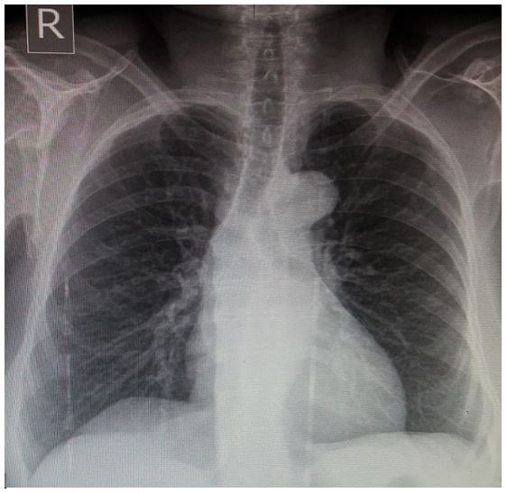
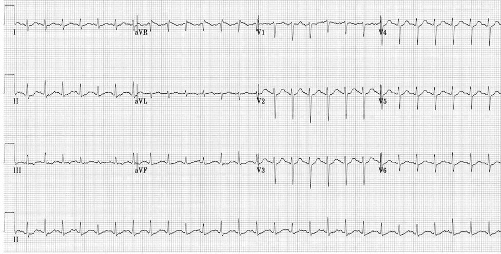
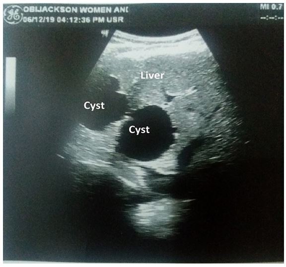
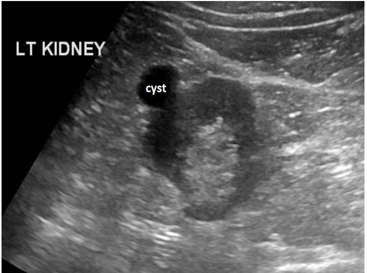
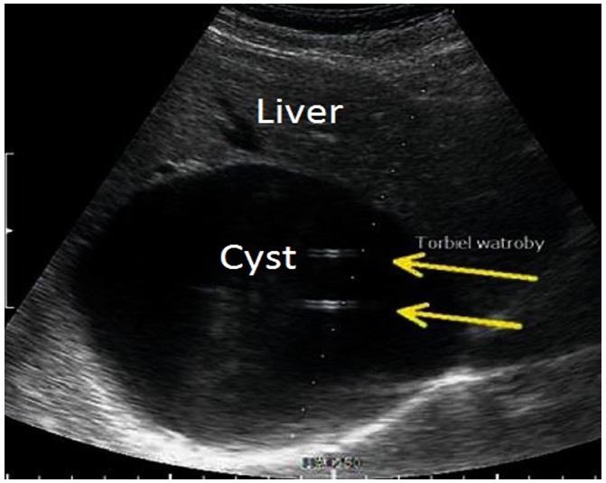
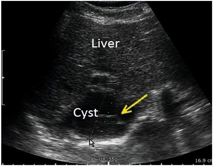
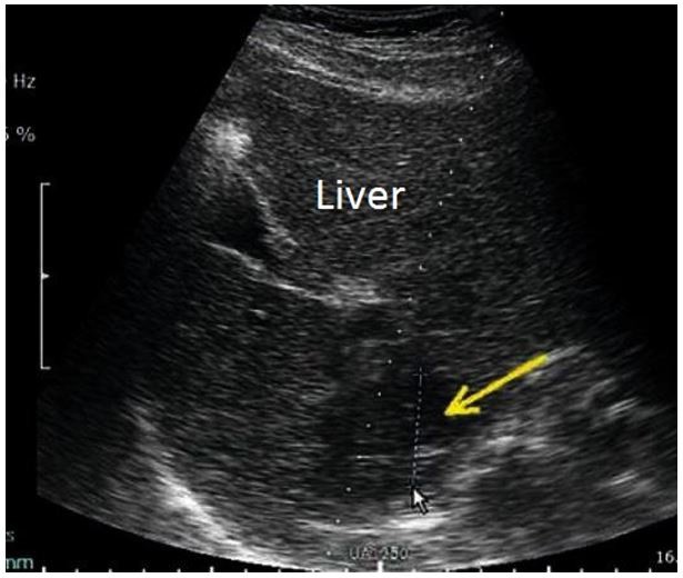
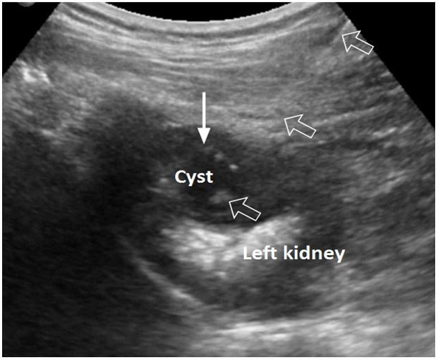
A 72 year old woman; an out-patient woman from South Eastern Nigeria presented to the radiology department for radio-diagnostic investigations; chest x-ray and abdomino-pelvic ultrasound. Her radiological request form showed a clinical history of general weakness, chest pain, palpitation, cough with purulent sputum and abdominal discomfort. Her BP was 180/110 mmHg and heart rate of 100 beats per minute. She presented in a stable general condition, except for blur vision. She had no history of smoking and diabetes mellitus. She had no other relevant diagnoses best known to our knowledge. She came with her daughter and grand-child. A chest x-ray was performed on her using General Electric (GE) Optima XR220amx mobile x-ray machine. An Agfa 35 x 40cm CRMD4.OT digital cassettes was used to perform the procedure. A postero-anterior (PA) chest projection was done while the patient was in an erect position, on a chest stand. A stationary grid was used to increase image contrast. Radiographic image was processed using an Agfa CR 30-X Reader. An abdomino-pelvic ultrasonography was performed on her using a LOGIQ P6/P6PRO ultrasound machine (General Electric Company, Wisconsin, USA) and with a curvilinear probe of frequency 4MHz. The chest radiograph showed a normal heart size with a cardiothoracic ratio (CTR) of 50%. There was prominent aortic unfolding. The lung fields are clear. Right diaphragmatic hump was noted. The costophrenic sulci were free. Degenerative changes were noted in the thoracic spine. The chest radiographic features were suggestive of systemic hypertension and no active lung disease. (Figure 1). Electrocardiography was also performed on her, which showed sinus tachycardia with no acute ST-T wave changes (Figure 2). The ultrasound showed a liver size of 15.6cm in craniocaudal span, measured at the mid-clavicular line. The liver showed a smooth outline. There were multiple, anechoic and thin wall rounded cysts of varying sizes located within the right and left lobes of the liver (segments III, IVb, VI and VII respectively). There were no echogenic debris or low level internal echoes noted in the cysts. The largest of the cysts which was located in segment VII measured 4.4cm. The background liver parenchyma showed a homogeneous echo-pattern. There was no ductal dilatation seen (Figure 3). The left kidney showed an upper moiety cortical cyst, which measured about 1.37cm in widest dimension (Figure 4). Both kidneys showed good sino-parenchymal differentiation and normal central sinus echoes. There were no hydronephrosis seen. There were no ascites. The ultrasound features were suggestive multiple hepatic Cysts and left upper pole simple cortical cyst.
The percutaneous drainage procedure was done by the patient’s physician with the radiologist’s assistance. The drainage utilized typical sets of drains with the diameter of 8F, mainly of a pigtail type. Drains were assembled using a suction pad to ultrasound transducer, following puncture with a puncture needle, along with the application of a soft guide. The procedure was performed under local anesthesia in combination with sedation. The needle was inserted through the lower intercostal spaces and as well below the costal arch for abdominal cavity assessment. The whole process of puncture, inserting the drainage and content removal by suction, along with the internal injection of 10% sodium chloride solution to prevent the possible reoccurrence of the cysts was monitored by ultrasound (Figure 5,6 and 7). Antibiotics regimens (ciprofloxacin) were administered orally (500 mg twice daily for 7 days) to the patient after the procedure in other to prevent or treat any possible infection. The patient symptoms were alleviated after the procedure. Her blood pressure reduced to 130/87mmHg. The patient is currently undergoing antihypertensive treatment regimens. Ultrasound guided percutaneous sclerotherapy of the left simple renal cyst was done using the same 10% sodium chloride solution (hypertonic saline) (Figure 8).
Systemic hypertension is a major risk factor for cardiovascular disease which occurs in 60% of patients with peripheral arterial disease, in 69% of patients with a first myocardial infarction, in 74% of patients with chronic cardiac failure and 77% of patients with a first stroke [9]. Reports from the 2013 World Health Day global brief on hypertension shows that Africa is worst hit [10].
Simple hepatic cysts are incidental radiological findings. These cysts may be classified as parasitic and non-parasitic. They are usually asymptomatic, but may be symptomatic due to local compression [11]. One of these compressive complications has been linked cause to edema, as a result of caval compression [11]. Our study showed that multiple liver cysts were noted in segments VI and VII, which appeared to be much closer to the inferior vena cava. The largest of the cysts was seen pressing on the inferior vena cava slightly. A Study described right sided heart failure [12] as a result of liver cyst compressing the right atrium [5]. The presented patient’s blood pressure reduction was in line with the case report of successive amelioration of hypertension after post-surgical removal of the cysts [13]..
Simple renal cysts were found to be related to hypertension [14,15]. This is because; blood pressure was discovered to have normalized after the surgical removal of cysts in hypertensive patients with simple renal cysts [16,17]. The largest diameter of the renal cyst in our report was 1.37cm. This is in line with the study which reported that there is high risk of hypertension if the largest diameter of the renal cyst appears to be more than 1cm [18]. A study showed that hypertension associated with simple renal cysts is likely to disappear after treatment. 46.1% of hypertensive patients who benefited from treatment had blood pressure normalization [19]. This is in line with our report, in which the patient’s blood pressure reduced after ultrasound guided percutaneous drainage of the cysts.
If the relationship between multiple hepatic cysts, simple renal cyst and hypertension is clarified through the suggested parameters and with supported literatures, increased risk of hypertension could be taken into serious consideration through observation, management and treatment of the ultrasound incidental findings of hepatic cysts and simple renal cyst in a patient with hypertension.
We are thankful to the head and staff of radiology department of Obijackson Women & Children Hospital for their support in the diagnostic investigations of the presented patient.