Ultrasound Guided Delineation of the Recurrent Laryngeal Nerve and the Tubercle of Zuckerkandl
Objective: To develop a direct, non-invasive method for the delineation of the Recurrent Laryngeal Nerve (RLN) and the Tubercle of Zuckerkandl (TZ).
Methods: Ultrasound imaging is performed on both sides of the neck in a study group of 50 persons without any prevailing medical conditions. RLN is identified on both sides and its distance is calculated from the Skin, Sternocleidomastoid muscle, midpoint of the trachea and the Carotid artery. Nerve diameter is also calculated. The TZ is searched for in the posterolateral aspect of the thyroid lobe.
Results: Distance of Left and Right RLN from Skin is 14.67±1.60mm and 14.42±1.88mm, from Sternocleidomastoid muscle is 8.36±0.37mm and 8.77±0.53mm, from carotid artery is 12.25±0.53mm and 12.71±0.37mm, from midpoint of the trachea is 7.92±0.3mm and 8.04±0.20mm. Diameter of the Left RLN and Right RLN is 1.13±0.14mm and 0.86±0.1 respectively. Another interesting finding is the TZ in the Study group persons.
Conclusion: This Study represents an initial attempt to evaluate and localize the RLN and TZ by means of ultrasound imaging. This direct and non-invasive method of assessment would prove to be beneficial in surgical practice.
Keywords: Recurrent Laryngeal Nerve; Tubercle of Zuckerkandl; Ultrasound
This study is focussed on the delineation of the Recurrent laryngeal nerve (RLN) and the Tubercle of Zuckerkandl (TZ) [1,2]. This research’s significance lies on numerous aspects.
Firstly, in the current surgical setting, minimally invasive surgery of the head and neck region is on a rise. Majority of the patients prefer to get minimally invasive surgery in comparison to the exhaustive surgeries which were performed few years back [3]. The main reason for this being involvement of much less health risk and better surgical outcomes. Although this surgery seems to encompass very less risk, there is a serious limitation. In the course of the surgical procedures, both the RLN and the TZ is quite vulnerable to damage. Secondly, Normal Thyroid and parathyroid surgeries are also likely to cause damage to the RLN and the TZ [4]. Finally, Radiofrequency ablation of thyroid nodules which is considered a common procedure nowadays in clinical settings, also involves the risk of endangering both the RLN and the TZ. Therefore, before performing any of the above mentioned surgical procedures, it is important to identify and localize the RLN as well as the TZ [5].
The RLN which branches off from the vagus nerve, provides the motor and sensory function to the larynx [6]. While ascending up the neck, as RLN produces more branches on the left than on the right side, it becomes easy to get identified on imaging studies. During thyroid surgery, RLN’s relatively medial location makes it susceptible to damage. One of the significant complications in the post thyroid and parathyroid surgical period is RLN palsy. Since 1970s, Intraoperative nerve monitoring (IONM) which has been used in head and neck surgeries has been a controversial subject with respect to its usefulness. Besides being expensive and less feasible in most situations, the results obtained are partially inaccurate.
So, our study is focussed on using ultrasound for the imaging of RLN. Being inexpensive and non-invasive, ultrasound can be easily performed before, during or after surgery for this nerve’s evaluation. The RLN, being vulnerable to injuries, is a significant nerve to be dealt with carefully during Minimally invasive head and neck surgeries, Thyroid or Parathyroid surgical procedures and also during Radiofrequency ablation of the thyroid nodules. Therefore, in this Research, with the utilization of ultrasound, the anomalies pertaining to RLN noticed in surgical scenarios can be reduced to a minimal level as a convenient visibility of the RLN is possible.
One more aspect of this study is the delineation of the TZ which anatomist Emil Zuckerkandl first described in 1902 [2]. Although it is a prominent anatomical entity, yet it was not a very familiar name till date. The significance of the TZ lies in the fact that, if not correctly identified and removed during thyroid surgery, it might prove to be a source of persistently present unrelieved symptoms or reoccurrence. But initial visibility is of concern. However a rare and moderately high risk situation is, when the RLN running laterally to an enlarged TZ, places it at an increased risk of damage during surgical dissection.
So, in this study, we identified the TZ by means of ultrasound [1]. During surgical procedures, this would prove to be beneficial due to easy and early identification of the RLN.
A study group of 50 healthy persons, 28 Female and 22 Male in the age group of 18-50 were chosen for carrying out this research study. These persons had neither been previously diagnosed with any medical conditions nor have they undergone any past surgeries. This human study carried out from January 2018 to August 2018, was performed in accordance with the Declaration of Helsinki and was approved by the Basic Research Program and Science Foundation of the Affiliated Traditional Chinese Medicine Hospital of Southwest Medical University, China. All adult participants provided verbal consent to participate in this study which was conducted during routine Ultrasound examination of the Thyroid/Parathyroid glands. After obtaining consent, diagnostic ultrasound was performed on each of the study group persons [1].
Ultrasound Imaging (using Model: IU22, Philips Company) of everyone’s neck on both left and right side were performed. To ensure better visualization, the patients were asked to lie down on their back while extending their neck over a pillow placed below their shoulders and then a considerable amount of coupling gel was applied on their neck. The linear probe used was of 5-12MHz. To obtain the best images in the neck, gain was varied. Both longitudinal and transverse plane scans were performed and it was necessary to rotate the head on each sides to obtain clear vascular images during the examination.
The RLN being located on both left and right side of the neck, its distance is then measured from the Skin, Sternocleidomastoid muscle, Carotid artery and the midpoint of the Trachea respectively. This was achieved by keeping the transducer in a transverse plane. Furthermore, the diameter of the RLN on both the sides is measured by keeping the transducer in a horizontal plane. Additionally, The TZ is looked for on the thyroid lobe’s posterolateral aspect.
Statistical analysis: Results were provided as mean ± SD. For the statistical analysis, T-tests were used. The calculations were done utilizing EXCEL software; with p<0.05 being considered significant.
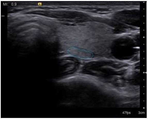
Upon performing Ultrasound imaging on both left and right side of the Neck, small neurovascular bundles were seen inside the tracheoesophageal groove. This structure being hypoechoic is surrounded by hyperechoic tissue (Figure 1). Magnified imaging is obtained for better results.
On the Left, RLN’s distance from Skin is 14.67±1.60mm, from Sternocleidomastoid muscle is 8.36±0.37mm, from carotid artery is 12.25±0.53mm and from the midpoint of the trachea is 7.92±0.3mm (Figure 2). Left RLN’s diameter is 1.13±0.14mm (Figure 3).
On the Right, RLN’s distance from Skin is 14.42±1.88mm, from Sternocleidomastoid muscle is 8.77±0.53mm, from carotid artery is 12.71±0.37mm, and from the midpoint of the trachea is 8.04±0.20mm. Right RLN’s diameter is 0.86±0.1 (Figure 4).
Identification of the TZ in the Study group persons is another interesting finding. Presenting as a postero-lateral extension of the thyroid gland, TZ was particularly noticed on the left side of the neck. This extended part of the thyroid lobe is identified and detected as the TZ on the basis of anatomical considerations (Figure 5).
With this study, a novel method for the convenient identification and localization of the RLN has been presented. Compared to other methods used in the past, this is direct, non-invasive, cheap and convenient [7].
After performing ultrasound imaging on both sides of the neck, small nerve bundles constituting hypoechoic structure surrounded by hyperechoic structure, were visible within the tracheoesophageal groove.
All the results, including measurements obtained, were in near accordance with the Gold Standard of delineation of the RLN and TZ which is the Post Mortem Studies on Cadavers [1,8,9].
Convenient delineation of the RLN by means of Ultrasound would prove to be quite beneficial for surgical practices in the recent scenario [4,6].Till current day, whenever the Surgeons performed Head or neck’s minimally invasive surgery, Thyroid / Parathyroid gland’s surgeries, or the radiofrequency ablation for the thyroid nodules, there existed a grave possibility of endangering the RLN [5,10,11]. Although Intraoperative Nerve monitoring performed by the Surgeons yielded successful results, it was an expensive method. Furthermore, if the Surgeons wished to get an idea regarding the structural and functional components of the RLN prior to surgery, they had to solely depend on expensive imaging modalities like MRI. This holds true for IONM as well [12]. One of the most important things to consider would be the follow-up period of the Surgeries. Most of the times, RLN developed anomalies during the post surgical period. Therefore, for all of the probable wrong outcomes, a cheap, direct, non-invasive and convenient to use modality was necessary, thereby bringing in the Ultrasound Imaging into picture. It’s understood that being a cheap and an easy to use imaging technology, Ultrasound if performed carefully, would have a high probability of acquiring best results at delineating the RLN during pre-operative, intra-operative or post operative periods.
There are many situations, where it has been noticed that if the RLN is not precisely identified before commencing with the surgery, then there would be a more likely chance of endangering the nerve. RLN being an important nerve in our body, is required for a precise vocal cord functioning. Post Thyroid or Parathyroid Surgeries, patients seem to suffer from hoarseness of voice which arises as a result of the RLN palsy. So, Ultrasound Guided delineation of the RLN might prove to be effective in both reduction and elimination of the problems entailed during Surgeries.
One more instance where this method of delineating RLN can be of great value, is during the surgical follow-up periods. Many times it has been noticed, that because Post operatively the RLN is likely to suffer from paralysis, it can be monitored at equal intervals to look for any anomalies.
An additional point of interest which came up during our study was the identification of the TZ. If mistakenly removed during thyroid surgery, it is significant as it might be a source of persistent unrelieved symptoms or reoccurrence. To undergo safe surgical dissection, a clear understanding of the TZ’s anatomy is quite crucial. With TZ being located lateral to the RLN, the nerve passes medial to it. If there is an early elevation of the TZ, that would enable easy and safe visualization of the RLN. But with the RLN running lateral to an enlarged TZ, it would be at an increased risk of getting damaged during surgery.
Hence, with ultrasound guided identification of the TZ being possible, it would be valuable in Medical practices all around, since this would also aid with the reduction and elimination of the chances of endangering RLN [13].
Ultrasound imaging, that has been presented as a novel method for the delineation of the RLN, is direct, non-invasive, convenient and cheap [7]. Delineation of the TZ is of additional benefit as it would help to avoid nerve anomalies [14].
This study is partially supported by the Basic Research Program and Science Foundation of The Affiliated Traditional Chinese Medicine Hospital of Southwest Medical University, China. The authors declare no funding and no conflict of interest for this article.


