Use Trichoscopy to Recognize Pili Annulati
Background:Pili annulati is a rare hair shaft abnormality, associated with an unknown pathogenesis that produces a macroscopic beading effect. An association with alopecia areata has been reported.
Cases presentation: In this report, we present the case of two girls, a 10-year-old girl and a 12-year-old girl, presenting for alopecia areata, with fortuitous discovery during the trichoscopy of the rest of the hair shaft of an alternation of light bands and dark bands evoking the diagnosis of pili annulati, confirmed by the polariscopic examination. The clinical and trichoscopic examination of the mother’s hair revealed the same aspect of alternating light and dark bands.
Conclusion: This abnormality of the hair, called pili annulati or ringed hair disease is a very rare pathology of the hair shaft. The clinical expression is heterogeneous within the same patient, 20 to 80% of the hair can be affected. Trichoscopy remains a non-invasive tool to suspect this disorder, confirmed by the identification of periodic light and dark bands on polariscopic examination. The pathogenesis is not yet clear. Finally, pili annulati has sometimes been observed in association with immunological disorders including alopecia areata, as in our patient.
Keywords:Pili Annulati; Hair Shaft Abnormality; Alopecia Areata; Trichoscopy
Pili annulati is a rare hair shaft abnormality of unknown pathogenesis which gives a gross beading effect. An association with alopecia areata has been reported. The management of diseases of the scalp has been enriched in recent years by a new tool: trichoscopy. This simple, fast, effective, non-invasive and inexpensive method greatly increases the dermatologist’s diagnostic performance with scalp diseases. We present the case of a patient who presents the combination of pili annulati and alopecia areata.
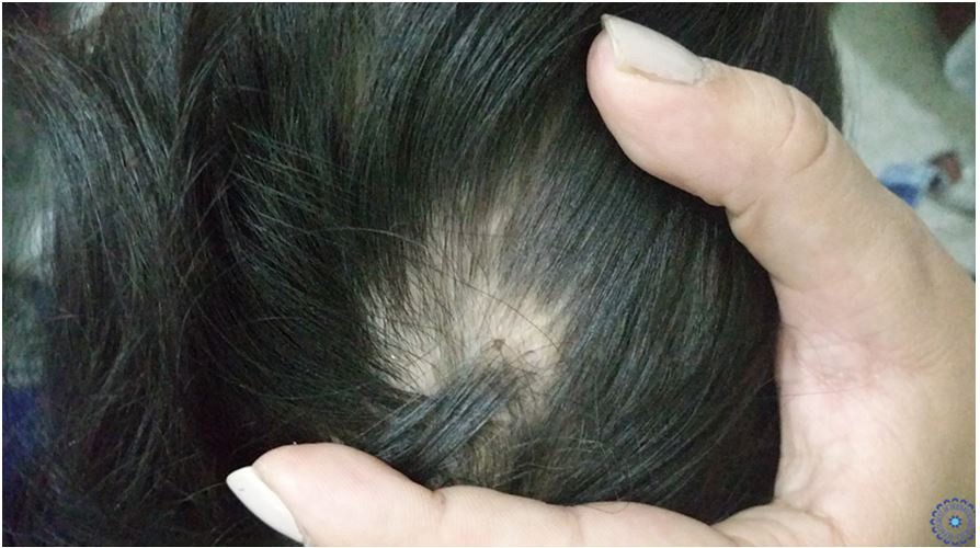
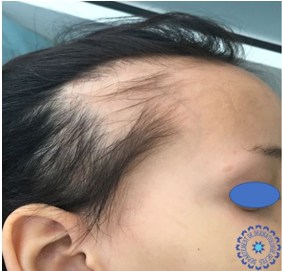
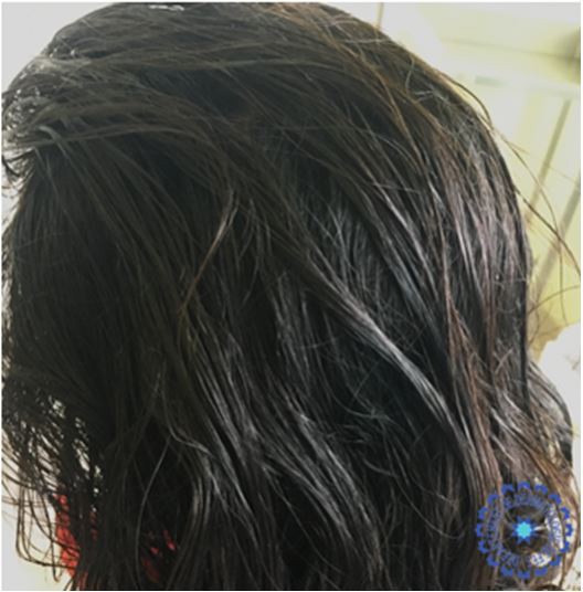
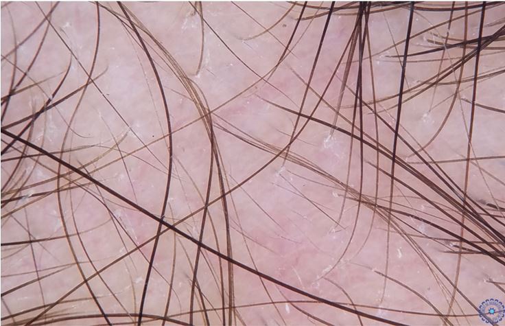
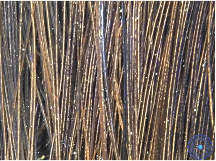
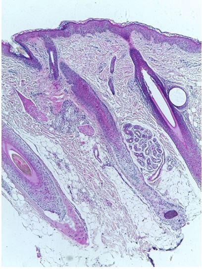
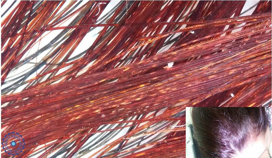
We report the case of two girls, a 10-year-old girl and a 12-year-old girl, presenting for alopecic plaques of the scalp. Having as a family history a similar symptomatology within their mother during childhood, treated by betamethasone injections with improvement. In whom the dermatological examination had objectified several non-cicatricial alopecic plates, a positive sign of traction, with shiny hair (Figure 1 and Figure 2). Trichoscopy of the alopecic plaques showed anisotrichy, coiled and fluffy hair with telangiectasias, and in the rest of the hair an alternation of light and dark bands typical of pili annulati (Figure 3, Figure 4 and Figure 5). The examination of the skin appendages had objectified an aspect of nail pitting of some nails of the hands (Figure 6 and Figure 7). A biopsy of the alopecic plaques was performed showing the presence in the dermis of some hair follicles miniaturized and other anagen type but with thin hair sheaths atrophic and having a very thin hair shaft incomplete incompletely cornified. This appearance of non-cicatricial alopecia is in relation with alopecia areata (Figure 8). The hair shaft examined under polarized light showed an alternation of dark and light areas at a regular interval over their entire length in favor of pili annulati. The child was treated with a very strong topical corticosteroid and Minoxidil 2%, with slight hair regrowth within the alopecic plaques (Figure 9). The assessment performed to detect an associated autoimmune pathology was normal.
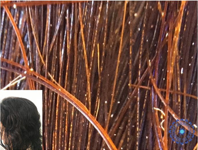
The clinical and trichoscopic examination of the mother’s hair revealed the same aspect of alternating light and dark bands (Figure 10 and Figure 11).
This abnormality of the hair, called pili annulati or ringed hair disease is a very rare pathology of the hair shaft. It was described for the first time in 1866 by Landois; almost 50 cases have been identified in the Anglo-Saxon literature [1]. Caucasian patients are the most affected [2]. The age of onset may vary from childhood to adulthood. The scalp is the most commonly affected, but other locations may also be involved such as pubic, beard or axillary area [3, 4]. The clinical expression is heterogeneous within the same patient, 20 to 80% of the hair can be affected. Trichoscopy remains a non-invasive tool to suspect this disorder, confirmed by the examination with polarized light [5, 6]. Although pili annulati is generally classified in hair abnormalities without capillary fragility, some patients report fragility induced by pathological cavities along the hair shaft [5]. The empty spaces reflect the light, giving the impression of rings [7]. There is no treatment for this disorder, but the prognosis is good and hair growth is usually normal [8, 9]. It is an autosomal dominant disease with variable expression; however, sporadic cases have also been described [9]. It is probably linked to a mutation on a single gene, not completely identified. In 2004, a gene locus responsible was mapped on chromosome 12q24.32-24.33. This anomaly is probably caused by a mutation in a regulatory gene that affects the proliferation or differentiation of follicular cells. The pathogenesis is not yet clear. Cortical cells, instead of containing tightly interwoven macrofibrils, would contain an insufficient amount of normal macrofibrillar material [10, 11]. Finally, pili annulati has sometimes been observed in association with immunological disorders including alopecia areata, as in our patient [3, 12, 13].
Pili annulati is an autosomal dominant disease affecting the hair [10, 11]. It is characterized by an alternation of dark and clear bands of the hair shaft better visualized by the dermoscope. Thus, we think that trichoscopy could replace the examination under polarized light for this disease. The concomitant existence of alopecia areata has been reported several times, but the direct association between these two pathologies seems unlikely. However, a systematic evaluation of patients with both diseases is needed to better characterize this association [4, 12-16].
What is known: Pili annulati is a rare hair shaft abnormality of unknown pathogenesis which gives a gross beading effect. An association with alopecia areata has been reported.
What is new: Trichoscopy remains a non-invasive tool to suspect this disorder, showing an alternation of light and dark bands.
The study has been approved by the ethics committee of faculty of medicine of fez.
An informed consent to participate in the study was obtained from the patient.
Written informed consent was obtained from the patient’s legal guardian(s) for publication of this case report and any accompanying images. A copy of the written consent is available for review by the Editor-in-Chief of this journal.
The authors declare that they have no conflicts of interest.
All the authors contributed for the interpretation of data for the work; and drafting the work or revising it critically for important intellectual content; and the final approval of the version to be published; and the agreement to be accountable for all aspects of the work in ensuring that questions related to the accuracy or integrity of any part of the work are appropriately investigated and resolved.