Reactive Amyloidosis in a Middle Aged Female with Behcet’s Disease – Case Report
Behcet’s disease is a multisystem vasculitis typically characterized by recurrent oral aphthae and genital ulcerations. Renal involvement in Behcet’s disease is less frequent and often less severe than in other types of vasculitis. We encountered a case of Reactive Amyloidosis secondary to Behcet’s disease in a middle aged female. Our patient had a partial remission in proteinuria after initiation of Prednisolone, Colchicine and Enalapril. Long term follows up is needed to assess the progression of the disease. This case is being reported for the rare and early occurrence of Renal Amyloidosis in a female with Behcet’s.
Keywords: Amyloidosis; Behcet’s disease; Oral aphthae; Reactive Amyloidosis; Prednisolone; Colchicine; Enalapril
Behçet’s disease, a vasculitis typically characterized by oro-genital aphthosis and systemic manifestations including ocular, cutaneous and neurological lesions. The clinical manifestations are due to angeitis and are remarkable for its ability to involve blood vessels of all sizes on both arterial and venous side of circulation. Behçet’s is diagnosed according to International criteria of Behçet’s Disease (ICBD). Renal disease in behçet’s is less frequent and often less severe than in other types of vasculitis. Patients with renal disease may have proteinuria, hematuria or renal insufficiency. The spectrum of renal diseases in behçet’s includes reactive amyloidosis (AA amyloidosis), renal artery aneurysms and interstitial nephritis. Patients with reactive amyloidosis present with nephrotic syndrome and mean duration from onset of behçet’s to amyloid nephropathy is 8 years. This case is being reported for the rare and early occurrence of renal amyloidosis in a female with severe behçet’s.
This 49-year-old female was admitted to our unit with fever, skin lesions and oral ulcers for a month. She had intermittent fever, painful pustular skin lesions from pin head to coin size over trunk and limbs and pedal edema of gradual onset. Active oral ulcers and healed vulval ulcers were noted. Her past medical history was significant for the occurrence of similar episodes of fever, skin lesions, oral and genital ulcers dating from November 2012. Labs revealed 3 + proteinuria and quantitative analysis showed a nephrotic range proteinuria (4 g/ d). Her complete hemogram and creatinine were unremarkable. Pathergy test was positive and Skin biopsy revealed evidence of Leucocytoclastic vasculitis.
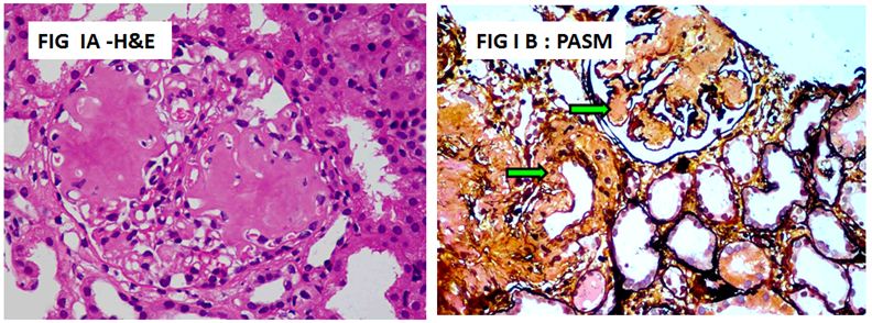
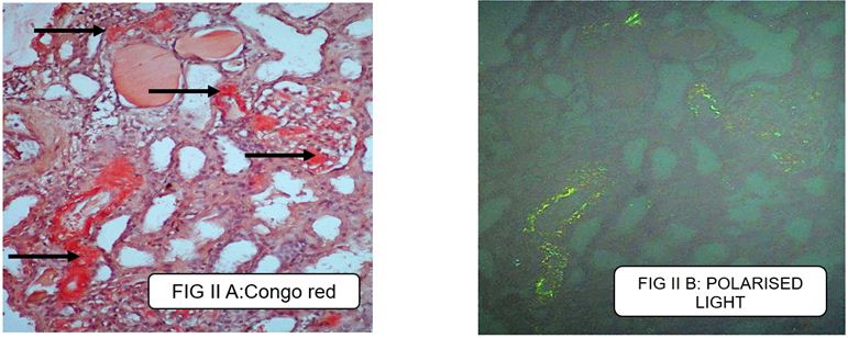
Renal Biopsy showed nodular mesangial expansion due to deposition of acellular amorphous material in light microscopic examination (Figure 1A). Special stains with PAS were strongly positive in bowman’s capsule and were weakly positive in the nodules. The nodules were silver negative compared to bowman’s capsule and tubular basement membrane (Figure 1B). The deposits took deep orange color on staining with congo red (Figure 2A) and exhibited apple green birefringence under polarized microscopy (Figure 2B). She was found to be HLA B 51 positive on further evaluation.
Behçet’s syndrome, multisystem angeitis is one of the rare diseases that lead, if not sufficiently treated, on to AA-Amyloidosis. Several case series of behçet’s with renal disease in males are noted. However, case reports of reactive amyloidosis in females with behçet’s is scattered while renal disease is not reported in literature. Hereby we report a case of severe behçet’s syndrome in a middle aged female with renal disease in the form of amyloidosis within 4 years of disease onset [1-3].
Behçet’s seems to cluster along the ancient silkroad extending from eastern Asia to the Mediterranean with prevalence being highest in Turkey and lower in European cohorts. The disease typically affects young adults between ages of 20 and 40 years. The sex predilection appears to have a geographical variability with male preponderance in the Mediterranean region while European and Japanese cohorts exhibiting female dominance. However, the severity of the disease including renal complications and mortality is uniformly high in males [4].
Our patient was a relatively older female with typical presentation of recurrent oral and genital aphthosis, cutaneous nodules and Pathergy. Ocular lesions which are seen in up to 75% of cases were not seen in our patient. The current criteria for Behçet’s diagnosis and classification are given by International Team for the Revision of the International Criteria for Behçet’s Disease (ITR-ICBD) which has higher sensitivity compared to the ISG criteria. Our patient satisfies the ICBD criteria [5].
Renal involvement in behçet’s includes proteinuria, hematuria or renal insufficiency. The spectrum of renal diseases in behçet’s was illustrated in a review of 159 patients by Akpolat et al. which showed AA amyloidosis in 69, glomerulonephritis in 51, vascular disease mostly renal artery aneurysms in 35 and interstitial nephritis in 4. Another series of 2007 Behçet’s patients from Korea by Sung Bin Cho et al. noted hematuria to be significantly higher compared to proteinuria (29 vs. 1.4 %) and IgA Nephropathy was the commonest renal lesion. Our patient presented with nephrotic range proteinuria and renal biopsy consistent with AA amyloidosis [6,7].
Patients with AA amyloidosis typically present with nephrotic syndrome or at least significant proteinuria with a male preponderance. In a case series of 14 patients, the mean time from behçet’s onset to amyloid nephropathy was 8 years with shorter duration in males. The presence of genital ulceration, thrombophlebitis, arthritis and neuropsychiatric involvement was more frequent among patients with amyloidosis than patients without amyloidosis. In our patient the time interval between onset of behçet’s and amyloidosis was much shorter (3¼ years) and she did not have any evidence of vascular, neurological or skeletal system involvement [8].
Amyloidosis is one of the prognostic factors affecting survival in Behçet’s disease. The 5-year survival rate in patients with Behçet’s and Reactive Amyloidosis is around 46 %. Corticosteroid is the corner stone of treatment for Behçet’s disease. Other immunosuppressive drugs being tried include cyclophosphamide, ciclosporine, methotrexate and Etanercept. Colchicine is widely used in the treatment of mucocutaneous manifestations and as an adjunct in the treatment of more serious manifestations [9].
Our patient was treated with prednisolone 30 mg/day, colchicine 1.2mg/day along with enalapril 5mg. She had partial remission of proteinuria (24-hour urinary protein – 1.3 gm) after 3 months with no further systemic flares and is currently on low dose prednisolone and maximally tolerated enalapril dosage of 10mg/d.