Sarcomatoid Carcinoma of Larynx: Case report
Sarcomatoid carcinoma is a rare form of laryngeal cancer which corresponds only 2-3% of all laryngeal cancers. This type of laryngeal cancer is presented mostly with hoarseness and dyspnea. On the endoscopic view, it can be detected with polypoid mass on the vocal cord and mixed with benign laryngeal polyps. But the behaviour and presentation of the cancer can be differed from other benign and malign tumors and may be very aggressive. The most effective treatment modality is not defined definitely. We represent a sarcomatoid cancer, presented with highly aggressive character, managed with total laryngectomy.
Keywords: Sarcomatoid carcinoma; Carcinosarcoma; Larynx Cancer; Head and Neck Cancer
Sarcomatoid carcinoma is a rare form of squamous cell carcinoma of larynx which also forms 2-3% of all laryngeal cancers [1-4]. This cancer is a variant of squamous cell cancer but its clinical behavior is more aggressive and more malignant in nature. Sarcomatoid carcinoma is composed of two components: sarcomatoid and epithelial cells which make the tumor biphasic. Most of the tumors are polypoid or pedunculated in clinical examination and usually cause obstructive respiratory symptoms in early stage of the disease. Pathological examination of biopsy material leads to diagnosis.
We represent a patient with sarcomatoid carcinoma who had a history of polypectomy and admitted to our clinic with new onset hoarseness and dyspnea.
A 44-years old male who was smoking for 30 years, admitted to our clinic with complaints of hoarseness and dyspnea. He had a history of vocal cord polyp excision approximately one month ago in another clinic. The pathological examination of surgical specimen revealed “benign polyp” then. He reported having similar symptoms before this surgery. Rigid and flexible endoscopic laryngeal examination showed a 2x1cm in size polypoid mass on left anterior 1/3 of vocal cord extending to the anterior comissure. The left vocal cord has paralyzed.
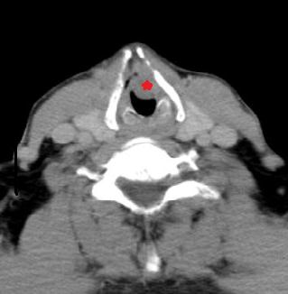
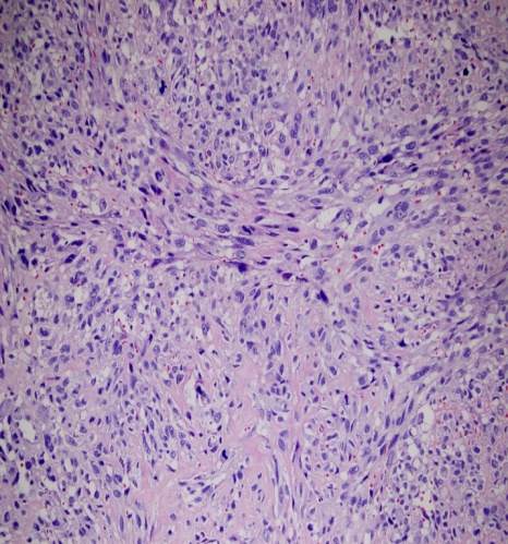
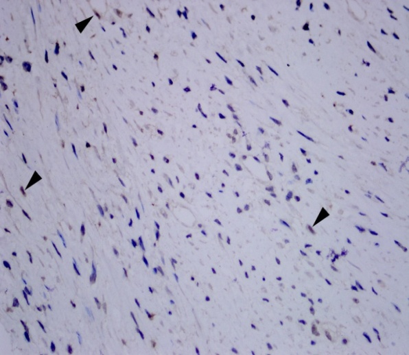
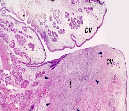
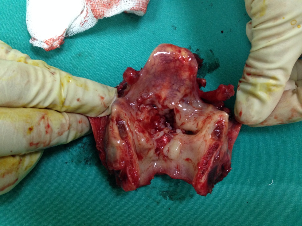
Computerized tomography examination of the larynx revealed a huge polypoid mass lesion on the left vocal fold (Figure 1). The lesion also destructed the thyroid cartilage and extended nearly 5 mm to subglottic area. We considered direct laryngoscopy and biopsy under general anesthesia. Urgent tracheostomy was done because of the mass obstructed the patient’s airway shortly before intubation. Biopsy material was sent to the pathological examination. The lesion is stained with cytokeratin, EMA, p63 dye. The tumor cells were spindle shaped in character with remarkable pleomorphism, atypical and intensive mitoses and the result was reported as sarcomatoid carcinoma of the larynx (Figure 2a,b,c,d). Due to T4 stage of the tumor and its malignant behavior, patient was operated with total laryngectomy and bilateral lateral neck dissection (Figure 3).
Squamous cell carcinoma is the most common type of laryngeal cancers [1]. Sarcomatoid carcinoma is highly malignant and rare form of squamous cell carcinoma. The incidence is 2-3% of all laryngeal cancer [5]. It is a biphasic tumor composed of epithelial and sarcomatoid parts [5]. The other synonyms of the sarcomatoid carcinoma is spindle cell carcinoma, pseudosarcoma and carcinosarcoma.
Sarcomatoid carcinoma is also believed to be monoclonal epithelial neoplasm with the sarcomatoid component derived from squamous epithelium during divergent mesenchymal differentiation [5].
The real cause is not exactly known but is mostly associated with cigarette smoking and alcohol consumption. In our case he had a 30 years history of cigarette smoking. Radiation exposure is also blamed but there is no definite information about the duration and dose of exposure [6-8].
Sarcomatoid carcinoma is 12 times more common in male and mostly seen in the 6th and 7th decades of life [4].
Clinically sarcomatoid carcinoma affects more commonly the glottic area. Therefore the patient’s chief complaints are hoarseness, dyspnea, cough and dysphagia [1]. These symptoms and location of the tumor help the clinician to diagnose the cancer at early stage.
The exact diagnosis is made by pathologic examination. Pathological examination must involve both squamous cell components and spindle shaped sarcomatous appearances. Without the characteristic cells we can not diagnose the sarcomotid carcinoma. Sarcomatoid carcinoma contains histologically squamous cell carcinoma with spindle cell changes that stimulate a sarcoma, in which the epithelial component is positive for Cytokeratin and sarcomatoid component is positive for Cytokeratin and occasionally positive for vimentin. Also on the pathological examination, spindle shaped tumor cells were stained with cytokeratin, EMA and p63 immunohistochemically. The cells are highly mitotic in appearence and polymorphic and strongly atypical in nature [12]. In our case the characteristics stains with spindle, highly mitotic and atipical nature of the cells are detected. These specialized features helped us to diagnose the sarcomatoid carcinoma.
Treatment modality depends on the location and stage of the tumor. Majority of the cases are diagnosed in stages T1 and T2 and have a good prognosis. This type of early stage tumors resected and radiation or chemoraditon treatment applied. But radiation therapy is not suggested because of the mesenchymal tumors are highly resistant to radiation. The late stage of tumors and the tumor located on transglottic and anterior commissure are surgically treated via total laryngectomy. But there is no concensus on the treatment modality in sarcomatoid carcinoma yet [9-12]. Local recurrence rate is reported between 16 and 32% according to the previous studies [13,14]. Incidence of cervical nodal metastasis is reported between 7,5 and 26% [4,13,15]. According to the Lambert et al, the distant metastasis to the lung and soft tissue 5% (15). All this data show us the highly malignant potential of the tumor behaviour. According to the our case, the patient’s polypoid mass was excised by surgically but within two months the lesion progressively grew and caused obstructive symptoms. This case prove that we must consider a radical treatment for the definite disease control on sarcomatoid carcinoma. The lesion of the patient was located on anterior comissure and tumor was transglottic lesion, so we carried out total laryngectomy and bilateral lateral neck disection successfully. 5 years survival is changed among 65-95% [4]. Our case is disease free according to 1 year follow up.
Sarcomatoid carcinoma is a rare tumor, variant of squamous cell carcinoma. It is characterized by pedunculated and polypoid mass on medical examination. The definite diagnosis is made via biopsy. Treatment method is controversial and many choices are used such as radiation chemotherapy, surgery and their combination. In our belief, we must consider the treatment modality according to the stage of the lesion. Surgery must be the mainstay of the treatment choice because of the mesenchymal nature of the tumor is resistant to radiation therapy.